Allantois
| Allantois | |
|---|---|
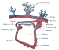
 Diagram illustrating a chicken egg in its 9th day with all extraembryonic membranes | |
 Sectional plan of the gravid human uterus in the third and fourth months of pregnancy | |
| Details | |
| Pronunciation | /əˈlæntɔɪs/ |
| Days | 16 |
| Identifiers | |
| Latin | Aallantois |
| MeSH | D000482 |
| TE | E6.0.1.2.0.0.2 |
| Anatomical terminology | |
The allantois (plural allantoides or allantoises) is a hollow sac-like structure filled with clear fluid that forms part of a developing amniote's conceptus (which consists of all embryonic and extra-embryonic tissues). It helps the embryo exchange gases and handle liquid waste.
The allantois, along with the amnion and chorion (other extraembryonic membranes), identify humans and other mammals as well as reptiles (including birds) as amniotes. Of the vertebrates, only the anamniotes (amphibians and non-tetrapod fish) lack this structure.
Function[]
This sac-like structure, whose name is the New Latin equivalent of "sausage" (in reference to its shape when first formed)[1] is primarily involved in nutrition and excretion, and is webbed with blood vessels. The function of the allantois is to collect liquid waste from the embryo, as well as to exchange gases used by the embryo.
In reptiles, birds, and monotremes[]
The structure first evolved in reptiles and birds as a reservoir for nitrogenous waste, and also as a means for oxygenation of the embryo. Oxygen is absorbed by the allantois through the egg shell.
In marsupials[]
In most marsupials, the allantois is avascular, having no blood vessels, but still serves the purpose of storing nitrogenous (NH3) waste. Also, most marsupial allantoises do not fuse with the chorion. An exception is the allantois of the bandicoot, which has a vasculature, and fuses with the chorion.
In placental mammals[]
In placental mammals, the allantois is part of and forms an axis for the development of the umbilical cord.
- The mouse allantois consists of mesodermal tissue, which undergoes vasculogenesis to form the mature umbilical artery and vein.[2]
- The human allantois is a caudal out-pouching of the yolk sac, which becomes surrounded by the mesodermal connecting stalk known as the body-stalk. The vasculature of the body-stalk develops into umbilical arteries that carry deoxygenated blood to the placenta.[3] It is externally continuous with the proctodeum and internally continuous with the cloaca. The embryonic allantois becomes the fetal urachus, which connects the fetal bladder (developed from cloaca) to the yolk sac. The urachus removes nitrogenous waste from the fetal bladder.[4]
Clinical significance[]
During the third week of development, the allantois protrudes into the area of the urogenital sinus. Between the 5th and 7th week of development, the allantois will become the urachus, a duct between the bladder and the yolk sac. A patent allantois can result in urachal cyst.
Additional images[]
Section through the embryo.

Diagram showing later stage of allantoic development with commencing constriction of the yolk-sac.
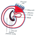
Diagram showing the expansion of amnion and delimitation of the umbilicus.

Model of human embryo 1.3 mm. long.

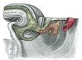
Tail end of human embryo from fifteen to eighteen days old.
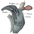
Cloaca of human embryo from twenty-five to twenty-seven days old.

Tail end of human embryo twenty-five to twenty-nine days old.
Tail end of human embryo thirty-two to thirty-three days old.

Opened uterus with cat fetus in midgestation: 1 umbilicus, 2 amniotic sac (chorion and amnion), 3 allantois, 4 Yolk sac, 5 developing marginal hematoma, 6 maternal part of placenta (endometrium)

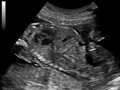
Ultrasound of fetus showing urachus duct from bladder to the umbilicus.

Ultrasound of fetus showing urachus duct from bladder to the umbilicus.

Ultrasound of fetus showing urachus duct from bladder to the umbilicus.
Ultrasound of fetus showing urachus duct from bladder to the umbilicus.

Ultrasound of fetus showing urachus duct from bladder to the umbilicus.


References[]
- ^ "Allantois". dictionary.com.
- ^ Downs, KM (1998). "The Murine Allantois". Curr Top Dev Biol. 39: 1–33. doi:10.1016/s0070-2153(08)60451-2. PMID 9475996.
- ^ Moore, Keith; Persaud, T.V.N.; Torchia, Mark G. (2016). The Developing Human: Clinically Oriented Embryology (10th ed.). Philadelphia: Elsevier. p. 58. ISBN 978-0-323-31338-4.
- ^ First AID for the USMLE Step 1 2008
External links[]
| Authority control |
|---|
- Embryology
- Animal reproductive system