Basophilic
This article does not cite any sources. (December 2009) |
Basophilic is a technical term used by pathologists. It describes the microscopic appearance of cells and tissues, as seen down the microscope, after a histological section has been stained with a basic dye. The most common such dye is haematoxylin.
Basophilic describes the appearance of structures seen in histological sections which take up basic dyes. The structures usually stained are those that contain negative charges, such as the phosphate backbone of DNA in the cell nucleus and ribosomes.
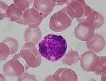
"Basophils" are cells that "love" the blue, and usually show up deep blue under standard staining techniques (H&E). Specifically, this term refers to:
- basophil granulocytes
- anterior pituitary basophils
Simplistically, acid pH stains (positive charge) are attracted to the base pH tissue (negative charge), so they are called "acidophilic stains". Eosinophils (basic components that like acids) are dyed red by the acid stain, eosin. "Basophils" (acid that like base components) are dyed blue by the basic stain, hematoxylin.
See also[]
- Histology