Bile duct hamartoma
| Bile duct hamartoma | |
|---|---|
 | |
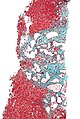
| Histopathology of a bile duct hamartoma, low magnification, H&E stain, showing a well demarcated lesion. |
A bile duct hamartoma or biliary hamartoma, is a benign tumour-like malformation of the liver.
They are classically associated with polycystic liver disease, as may be seen in the context of polycystic kidney disease, and represent a malformation of the liver plate.[1]
Cause[]
It is a developmental anomaly in which abnormal tissues are present at normal site. Due to failure of regression of embryonic billiary duct.[citation needed]
Diagnosis[]

- Small to medium sized, irregularly shaped bile ducts lined by bland cuboidal epithelium (may also be flattened).
- Prominent intervening collagenous stroma.
- Bile ducts containing eosinophilic debris (may also contain inspissated bile)
At CT scans, bile duct hamartomas appear as small, well-defined hypo- or isoattenuating masses with little or no enhancement after contrast administration.[3] At MRI, they appear hypointense on T1-weighted images, iso- or slightly hyperintense on T2-weighted images, and hypointense after administration of gadolinium based contrast-agent.[3] On imaging, multiple hamartomas may look similar to metastases or microabscesses.[citation needed]
Treatment[]
Observation as there is increased risk of cholangiocarcinoma.[citation needed]
Eponym[]
The eponymous terms (von Meyenburg complex, Meyenburg complex) are named for Hanns von Meyenburg.[4][5]
Additional images[]

Micrograph of a bile duct hamartoma. Trichrome stain. Intermediate magnification

Micrograph of a bile duct hamartoma. Trichrome stain, high magnification
Low magnification micrograph of a bile duct hamartoma. Trichrome stain.
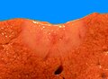
Gross pathologic appearance of a large bile duct hamartoma.


See also[]
References[]
- ^ Desmet, VJ (Jan 1998). "Ludwig symposium on biliary disorders--part I. Pathogenesis of ductal plate abnormalities". Mayo Clin Proc. 73 (1): 80–9. doi:10.4065/73.1.80. PMID 9443684.
- ^ Upasana Joneja, M.D. "Liver & intrahepatic bile ducts - Developmental anomalies / cysts - Von Meyenburg complex". Pathology Outlines. Topic Completed: 23 November 2020. Minor changes: 23 November 2020
- ^ Jump up to: a b Horton, KM; Bluemke, DA; Hruban, RH; Soyer, P; Fishman, EK (Mar–Apr 1999). "CT and MR imaging of benign hepatic and biliary tumors". Radiographics. 19 (2): 431–51. doi:10.1148/radiographics.19.2.g99mr04431. PMID 10194789.
- ^ synd/1693 at Who Named It?
- ^ H. von Meyenburg. Über die Zyztenleber. Beiträge zur pathologischen Anatomie und zur allgemeinen Pathologie, Jena, 1918, 64: 477-532.
- Congenital disorders of digestive system
- Neoplasm stubs