Canals of Hering
| Canals of Hering | |
|---|---|
 The canal of Hering leads to the | |
| Details | |
| Identifiers | |
| Latin | Ductulus bilifer |
| Anatomical terminology | |
The canals of Hering, or intrahepatic bile ductules, are part of the outflow system of exocrine bile product from the liver. Liver stem cells are located in the canals of Hering.
Structure[]
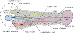
They are found between the bile canaliculi and interlobular bile ducts near the outer edge of a classic liver lobule.[1][2]
Histology[]
Histologically, the cells of the ductule are described as simple cuboidal epithelium, lined partially by cholangiocytes and hepatocytes. They may not be readily visible but can be differentially stained by cytokeratins CK19 and CK7.
Clinical relevance[]
The canals of Hering are destroyed early in primary biliary cholangitis and may be primary sites of scarring in methotrexate toxicity. Research has indicated the presence of intraorgan stem cells of the liver that can proliferate in disease states, so-called oval cells.[3]
History[]
They are named for Ewald Hering.[4]
References[]
- ^ Ross, M.H. & Pawlina, W. 2003. Histology: A Text and Atlas, 4th Edition. Lippincott Williams & Wilkins, Philadelphia.
- ^ Gartner, L.P. & Hiatt, J.L. 2000. Color Atlas of Histology, 3rd Edition. Lippincott Williams & Wilkins, Philadelphia.
- ^ Saxena, R. & Theise, N. 2004. Canals of Hering: Recent Insights and Current Knowledge, Semin Liver Dis 24: 43-48.
- ^ Hering E. Uber den Bau der Wirbelthierleber. Archiv für mikroskopische Anatomie 1867;3:88–118
- Hepatology