Carina of trachea
| Carina of trachea | |
|---|---|
 | |
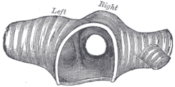 Transverse section of the trachea, just above its bifurcation, with a bird’s-eye view of the interior. (Carina not labeled; the ridge that separates the left and right bronchus.) | |
| Details | |
| System | Respiratory system |
| Identifiers | |
| Latin | Carina tracheae, bifurcatio tracheae |
| TA98 | A06.3.01.009 |
| TA2 | 3225 |
| FMA | 7465 |
| Anatomical terminology | |
In anatomy, the carina is a ridge of cartilage in the trachea that occurs between the division of the two main bronchi.[1][2]
Structure[]
The carina occurs at the lower end of the trachea (usually at the level of the 4th to 5th thoracic vertebra).[1][3] This is in line with the sternal angle, but the carina may raise or descend up to two vertebrae higher or lower with breathing. The carina lies to the left of the midline, and runs antero-posteriorly (front to back).
The bronchial arteries supply the carina and the rest of the lower trachea.[4]
The carina is around the area posterior to where the aortic arch crosses to the left of the trachea.[3] The azygos vein crosses right to the trachea above the carina.[5]
Clinical significance[]
Foreign bodies that fall down the trachea are more likely to enter the right bronchus.
The mucous membrane of the carina is the most sensitive area of the trachea and larynx for triggering a cough reflex.
Widening and distortion of the carina is a serious sign because it usually indicates carcinoma of the lymph nodes around the region where the trachea divides.
Tracheobronchial injury, an injury to the airways, occurs within 2.5 cm of the carina 60% of the time.[6]
Additional images[]

Anatomical dissection of trachea and main bronchi showing the carina

Anatomy of the trachea


References[]
- ^ a b Fernandez, Luis G.; Norwood, Scott H.; Berne, John D. (2008-01-01), Asensio, JUAN A.; Trunkey, DONALD D. (eds.), "Tracheal, Laryngeal, and Oropharyngeal Injuries", Current Therapy of Trauma and Surgical Critical Care, Philadelphia: Mosby, pp. 215–226, doi:10.1016/b978-0-323-04418-9.50036-9, ISBN 978-0-323-04418-9, retrieved 2020-11-17
- ^ Reynolds, Susan D.; Pinkerton, Kent E.; Mariassy, Andrew T. (2015-01-01), Parent, Richard A. (ed.), "Chapter 6 - Epithelial Cells of Trachea and Bronchi", Comparative Biology of the Normal Lung (Second Edition), San Diego: Academic Press, pp. 61–81, doi:10.1016/b978-0-12-404577-4.00006-0, ISBN 978-0-12-404577-4, retrieved 2020-11-17
- ^ a b Schipper, Paul; Sukumar, Mithran; Mayberry, John C. (2008-01-01), Asensio, JUAN A.; Trunkey, DONALD D. (eds.), "Pertinent Surgical Anatomy of the Thorax and Mediastinum", Current Therapy of Trauma and Surgical Critical Care, Philadelphia: Mosby, pp. 227–251, doi:10.1016/b978-0-323-04418-9.50037-0, ISBN 978-0-323-04418-9, retrieved 2020-11-17
- ^ Acocella, F.; Brizzola, S. (2011-01-01), Bosworth, Lucy A.; Downes, Sandra (eds.), "12 - Tracheal tissue regeneration", Electrospinning for Tissue Regeneration, Woodhead Publishing Series in Biomaterials, Woodhead Publishing, pp. 242–279, doi:10.1533/9780857092915.2.242, ISBN 978-1-84569-741-9, retrieved 2020-11-17
- ^ Hardie, E. (2014-01-01), Langley-Hobbs, Sorrel J.; Demetriou, Jackie L.; Ladlow, Jane F. (eds.), "Chapter 46 - Trachea and bronchus", Feline Soft Tissue and General Surgery, W.B. Saunders, pp. 531–540, doi:10.1016/b978-0-7020-4336-9.00046-9, ISBN 978-0-7020-4336-9, retrieved 2020-11-17
- ^ Chu CP, Chen PP (April 2002). "Tracheobronchial injury secondary to blunt chest trauma: Diagnosis and management". Anaesth Intensive Care. 30 (2): 145–52. doi:10.1177/0310057X0203000204. PMID 12002920.
External links[]
| Look up carina in Wiktionary, the free dictionary. |
- Atlas image: lung_carina at the University of Michigan Health System - "Cast of trachea and bronchi, anterior view" (#2)[dead link]
- "Trachea and carina — tomogram, coronal plane" at SUNY Downstate Medical Center Archived 2016-03-03 at the Wayback Machine
- Carina tracheae entry in the public domain NCI Dictionary of Cancer Terms
![]() This article incorporates public domain material from the U.S. National Cancer Institute document: "Dictionary of Cancer Terms".
This article incorporates public domain material from the U.S. National Cancer Institute document: "Dictionary of Cancer Terms".
Authority control | |
|---|---|
| Scientific databases | |
| Other | |
- Trachea