Cystohepatic triangle
| Cystohepatic triangle | |
|---|---|
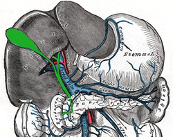
 The cystic artery branches from the right hepatic artery. | |
 Relationship to other vessels. | |
| Details | |
| Identifiers | |
| Latin | trigonum cystohepaticum |
| TA98 | A10.1.02.428 |
| TA2 | 3756 |
| FMA | 24230 |
| Anatomical terminology | |
The cystohepatic triangle (or hepatobiliary triangle) is an anatomic space bordered by the cystic duct inferiorly, the common hepatic duct medially, and the inferior surface of the liver superiorly. The cystic artery lies within the hepatobiliary triangle, which is used to locate it during a laparoscopic cholecystectomy.
Structure[]
The hepatobiliary triangle is the area bound by the:
- cystic duct inferiorly.[1][2]
- common hepatic duct medially.[1][2]
- inferior margin of the liver superiorly.[1][2]
It is covered in peritoneum both anteriorly and posteriorly.[2] It contains the cystic artery and cystic lymph nodes.[2] The right hepatic artery may also pass through the hepatobiliary triangle.[2]
Clinical significance[]
This section does not cite any sources. (January 2010) |
General surgeons frequently quiz medical students on this term and the name for the lymph node located within the triangle, Mascagni's lymph node or Lund's node, however many often erroneously refer to it as "Calot's node." The latter is frequently enlarged due to inflammation of the gallbladder (e.g. cholecystitis) or the biliary tract (e.g. cholangitis) and may be removed along with the gallbladder during surgical treatment (cholecystectomy).
The cystic artery lies within the hepatobiliary triangle, which is used to locate it during a laparoscopic cholecystectomy.[3][4] It may also contain an accessory right hepatic artery or an anomalous sectoral bile ducts. As a result, dissection in the triangle of Calot is ill-advised until the lateral-most structures have been cleared and identification of the cystic duct is definitive. According to SESAP 12 (produced and distributed by the American College of Surgeons) dissection in the triangle of Calot is the most common cause of common bile duct injuries.
History[]
Another name used to refer to the hepatobiliary triangle is Calot's triangle, after Jean-François Calot.[5][6] Calot's original description of the triangle in 1891 included the cystic duct, the common hepatic duct, and the cystic artery (not the inferior border of the liver as is commonly believed).[3]
References[]
- ^ a b c Schwartz's Manual of Surgery BRUNICARDI C.F 10th edition
- ^ a b c d e f Connor, Saxon J.; Perry, William; Nathanson, Leslie; Hugh, Thomas B.; Hugh, Thomas J. (May 2014). "Using a standardized method for laparoscopic cholecystectomy to create a concept operation-specific checklist". HPB. 16 (5): 422–429. doi:10.1111/hpb.12161. ISSN 1365-182X. PMC 4008160. PMID 23961737.
- ^ a b Haubrich, William (November 2002). "Calot of the triangle of Calot". Gastroenterology. 123 (5): 1440. doi:10.1053/gast.2002.1231440. PMID 12404217.
- ^ Orozco, Hector; Mercado, Miguel Angel; Takahashi, Takeshi; Hernández-Ortiz, Jorge; Capellán^S, Juan Félix; Garcia-Tsao, Guadalupe (June 1992). "Elective treatment of bleeding varices with the Sugiura operation over 10 years". The American Journal of Surgery. 163 (6): 585–589. doi:10.1016/0002-9610(92)90562-6. ISSN 0002-9610. PMID 1595838.
- ^ synd/4023 at Who Named It?
- ^ J. F. Calot. De la cholécystectomie. Doctoral thesis, Paris, 1891.
6. Bailey & Love's Short Practice of Surgery 26th edition (see page 1098).
| Authority control: Scientific databases |
|---|
- Abdomen
- Hepatology