Ferrichrome
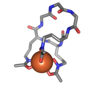 Ferrichrome (sticks) bound to an iron atom (orange)
| |
| Names | |
|---|---|
| IUPAC name
N-[3-[4,16-bis[3-[acetyl(oxido)amino]propyl]-2,5,8,11,14,17-hexaoxo-3,6,9,12,15,18-hexazacyclooctadec-1-yl]propyl]-N-oxidoacetamide; iron(3+)
| |
| Identifiers | |
| |
3D model (JSmol)
|
|
| ChemSpider | |
| ECHA InfoCard | 100.036.081 |
| EC Number |
|
PubChem CID
|
|
| UNII | |
CompTox Dashboard (EPA)
|
|
| Properties | |
| C27H42FeN9O12 | |
| Molar mass | 740.529 g·mol−1 |
Except where otherwise noted, data are given for materials in their standard state (at 25 °C [77 °F], 100 kPa). | |
| Infobox references | |
Ferrichrome is a cyclic hexa-peptide that forms a complex with iron atoms. It is a siderophore composed of three glycine and three modified ornithine residues with hydroxamate groups [-N(OH)C(=O)C-]. The 6 oxygen atoms from the three hydroxamate groups bind Fe(III) in near perfect octahedral coordination.
Ferrichrome was first isolated in 1952, has been found to be produced by fungi of the genera Aspergillus, Ustilago, and Penicillium.[1]
Biological function[]
Ferrichrome is a siderophore, which are metal chelating agents that have a low molecular mass and are produced by microorganisms and plants growing under low iron conditions. The main function of siderophores is to chelate ferric iron (Fe3+) from insoluble minerals from the environment and make it available for microbial and plant cells. Iron is important in biological functions as it acts as a catalyst in enzymatic processes, as well as for electron transfer, DNA and RNA synthesis, and oxygen metabolism.[2] Although iron is the fourth most abundant element in the earth’s crust,[3] bioavailability of iron in aerobic environments is low due to formation of insoluble ferric hydroxides. Under iron limitation, bacteria scavenge for ferric iron (Fe3+) by up-regulating the secretion of siderophores in order to meet their nutritional requirements.[4] Recent studies have shown that ferrichrome has been used as a tumor- suppressive molecule produced by L. casei. The study from the Department of Medicine and Asahikawa Medical University, suggests that ferrichrome has a greater tumor-suppressive effect than other drugs currently used to fight colon cancer, including cisplatin and 5-fluoro-uracil. Ferrichrome also had less of an effect on non-cancerous intestinal cells than the two previously mentioned cancer fighting drugs. It was determined that ferrichrome activated the C-Jun N-terminal kinases, which induced apoptosis. The induction of apoptosis by ferrichrome is reduced by the inhibition of the c-jun N-terminal kinase signaling pathway.[5]
Uptake[]
Iron is essential for the most important biological processes such as DNA and RNA synthesis, glycolysis, energy generation, nitrogen fixation and photosynthesis, therefore uptake of iron from the environment and transport into the organism are critical life processes for almost all organisms.[6] The problem is when environmental iron is exposed to oxygen it is mineralized to its insoluble ferric oxy hydroxide form which can not be transported into the cells and therefore is not available for use by the cell.[6] To overcome this, bacteria, fungi and some plants synthesize siderophores, and secrete it into an extracellular environment where binding of iron can occur.[6] It is important to note microbes make their own type of siderophore so that they are not competing with other organisms for iron uptake.[6] Ferrichrome is a unique siderophore, that is of the hydroxamate class (tris(hydroxamate)).[7] It has an exceptionally high binding affinity of logβ110 = 29.07 to ferric iron compared to [Fe(edta)]− that is logβ110 = 25.1 respectively. This indicates that it has an extremely high Fe3+ specificity and does not bind other metals in high concentration.[7] For example, saccharomyces cerevisiae is a species of yeast that can uptake the iron bound siderophore through transporters of the ARN family.[7] [Fe3+( siderophore)](n-3)- binds to a receptor-transporter on the cell surface and then is up taken.[7] The exact mechanism how iron enters the cell using these transporters is not understood, but it known that once it enters the cell it accumulates in the cytosol.[7] In saccharomyces cerevisiae, ferrichrome is specifically taken up by ARN1P as it has 2 binding sites and ferrichrome can the higher affinity site through endocytosis.[7] Ferrichrome chelates stay stable in the cell and allow for iron storage, but can be easily mobilized to meet the metabolic needs of the cell.[7]
Receptor[]
E. coli has a receptor protein called FhuA (ferric Hydroxamate).[8]
FhuA’s is an energy-coupled transporter and receptor.[8] It is a part of the integral outer membrane proteins and works alongside an energy transducing protein TonB.[9] It is involved in the uptake of iron in complex with ferrichrome by binding and transporting ferrichrome-iron across the cell’s outer membrane.[9]
The blue ribbons represent β-barrel wall that is 69Å long x 40-45Å diameter that represents the C-terminus residues. It has 22 antiparallel β strands. The yellow ribbon in the center is a “cork” which is a distinct domain for the N-terminus residues.[9]
FhuA has L4 strand and its role is to transport ferrichrome into the β-barrel wall. The ferrichrome complex then binds tightly to both the β-barrel wall and the "cork".[9] As a result, these binding triggers two key conformation changes to iron-ferrichrome complex to transfer energy to the cork. This energy transfer results in subsequent conformational changes that transport iron-ferrichrome to the periplasmic pocket which signal a ligand loaded status of the receptor.[9] These subtle shifts disrupt the binding of iron-ferrichrome to the cork which then allows the permeation of the ferrichrome-iron to a putative channel-forming region. The inner wall of the β-barrel provides a series of weak binding sites to pull ferrichrome along.[9] FhuD is a high affinity binding protein in the periplasmic pocket that also aids in unidirectional transport across the cell envelope.[9]
See also[]
References[]
- ^ Ferrichrome Archived 2010-01-13 at the Wayback Machine, Virtual Museum of Minerals and Molecules, University of Wisconsin
- ^ Ahmed E, Holmström SJ (May 2014). "Siderophores in environmental research: roles and applications". Microbial Biotechnology. 7 (3): 196–208. doi:10.1111/1751-7915.12117. PMC 3992016. PMID 24576157.
- ^ Loper JE, Buyer JS (September 1990). "Siderophores in Microbial Interactions on Plant Surfaces". Molecular Plant-Microbe Interactions. 4: 5–13. doi:10.1094/mpmi-4-005.
- ^ Chatterjee A, O'Brian MR (April 2018). "Rapid evolution of a bacterial iron acquisition system". Molecular Microbiology. 108 (1): 90–100. doi:10.1111/mmi.13918. PMC 5867251. PMID 29381237.
- ^ Konishi H, Fujiya M, Tanaka H, Ueno N, Moriichi K, Sasajima J, et al. (August 2016). "Probiotic-derived ferrichrome inhibits colon cancer progression via JNK-mediated apoptosis". Nature Communications. 7: 12365. doi:10.1038/ncomms12365. PMC 4987524. PMID 27507542.
- ^ Jump up to: a b c d Hannauer M, Barda Y, Mislin GL, Shanzer A, Schalk IJ (March 2010). "The ferrichrome uptake pathway in Pseudomonas aeruginosa involves an iron release mechanism with acylation of the siderophore and recycling of the modified desferrichrome". Journal of Bacteriology. 192 (5): 1212–20. doi:10.1128/JB.01539-09. PMC 2820845. PMID 20047910.
- ^ Jump up to: a b c d e f g Moore RE, Kim Y, Philpott CC (May 2003). "The mechanism of ferrichrome transport through Arn1p and its metabolism in Saccharomyces cerevisiae". Proceedings of the National Academy of Sciences of the United States of America. 100 (10): 5664–9. Bibcode:2003PNAS..100.5664M. doi:10.1073/pnas.1030323100. PMC 156258. PMID 12721368.
- ^ Jump up to: a b Braun V (June 2009). "FhuA (TonA), the career of a protein". Journal of Bacteriology. 191 (11): 3431–6. doi:10.1128/JB.00106-09. PMC 2681897. PMID 19329642.
- ^ Jump up to: a b c d e f g Ferguson AD, Hofmann E, Coulton JW, Diederichs K, Welte W (December 1998). "Siderophore-mediated iron transport: crystal structure of FhuA with bound lipopolysaccharide". Science. 282 (5397): 2215–20. Bibcode:1998Sci...282.2215F. doi:10.1126/science.282.5397.2215. PMID 9856937.
- Iron(III) compounds
- Peptides
- Siderophores