Hyperdontia
This article needs additional citations for verification. (March 2020) |
| Hyperdontia (Supernumerary Teeth) | |
|---|---|
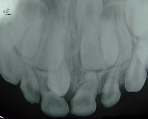 | |
| Supernumerary teeth in the premaxillary area | |
| Specialty | Dentistry |
| Symptoms | supernumerary teeth coming out from the gum or in the mouth |
| Complications | supernumerary teeth growing into the gum |
| Types | 5[citation needed] |
| Causes | Gardner's syndrome
Ehlers-Danlos syndrome Cleft palate Cleidocranial dysplasia Genetic disorder |
| Risk factors | supernumerary teeth causing problems in the dental arch |
| Differential diagnosis | Hypodontia |
| Treatment | Dental surgery |
Hyperdontia is the condition of having supernumerary teeth, or teeth that appear in addition to the regular number of teeth (32 in the average adult). They can appear in any area of the dental arch and can affect any dental organ. The opposite of hyperdontia is hypodontia, where there is a congenital lack of teeth, which is a condition seen more commonly than hyperdontia.[1] The scientific definition of hyperdontia is "any tooth or odontogenic structure that is formed from tooth germ in excess of usual number for any given region of the dental arch."[2] The additional teeth, which may be few or many, can occur on any place in the dental arch. Their arrangement may be symmetrical or non-symmetrical.
Signs and symptoms[]
The presence of a supernumerary tooth, particularly when seen in young children, is associated with a disturbance of the maxillary incisor region. This commonly results in the impaction of the incisors during the mixed dentition stage. The study debating this also considered many other factors such as: the patient's age, number, morphology, growth orientation and position of the supernumerary tooth. Alongside this issue, the presence of an extra tooth can impede the eruption of adjacent additional or normal teeth. Therefore, the presence of a supernumerary tooth when found must be approached with the appropriate treatment plan, incorporating the likelihood of incisal crowding.[3] In some individuals, the additional teeth can erupt far from the dental arch, within the maxillary sinus. The extra teeth may also migrate to a different location after development.[1] In some cases, supernumerary teeth can lead to the formation of cysts. Crowding is also frequently seen in people with hyperdontia.[2]
Causes[]
There is evidence of hereditary factors along with some evidence of environmental factors leading to this condition. While a single excess tooth is relatively common, multiple hyperdontia is rare in people with no other associated diseases or syndromes.[4] Many supernumerary teeth never erupt, but they may delay eruption of nearby teeth or cause other dental or orthodontic problems.[5][6] Molar-type extra teeth are the most common type. Dental X-rays are often used to diagnose hyperdontia.
It is suggested that supernumerary teeth develop from a third tooth bud arising from the dental lamina near the regular tooth bud or possibly from splitting the regular tooth bud itself. Supernumerary teeth in deciduous (baby) teeth are less common than in permanent teeth.
Related conditions[]
Hyperdontia is seen in a number of disorders, including Gardner's syndrome and cleidocranial dysostosis, where multiple supernumerary teeth are seen that are usually impacted.[citation needed]
Other associated conditions are: Cleidocranial dysplasia, Ehlers–Danlos syndrome Type III, Ellis–van Creveld syndrome, Gardner's syndrome, Goldenhar syndrome, Hallermann–Streiff syndrome, Orofaciodigital syndrome type I, Incontinentia pigmenti, Marfan syndrome, Nance–Horan syndrome, and Tricho-rhino-phalangeal syndrome Type 1.
Diagnosis[]
Supernumerary teeth may be detected by taking two different dental X-rays at different angles. Examples of this may be an intra-oral X-ray (one that is taken inside the mouth) and a panoramic radiograph. However, these X-rays are 2D and therefore do not accurately portray the 3D view of the teeth.[2]
Types[]

Supernumerary teeth can be classified by shape and by position. The shapes include the following:
- Supplemental (where the tooth has a normal shape for the teeth in that series);
- Tuberculate (also called barrel shaped);
- Conical (also called peg shaped);
- Compound odontoma (multiple small tooth-like forms);
- Complex odontoma (a disorganized mass of dental tissue)[7]
When classified by position, a supernumerary tooth may be referred to as a mesiodens, a paramolar, or a distomolar.[7] Occasionally, these teeth do not erupt into the oral cavity but manifest as a malocclusion.[8]
The most common supernumerary tooth is a mesiodens, which is a malformed, peg-like tooth that occurs between the maxillary central incisors.
Fourth and fifth molars that form behind the third molars are another kind of supernumerary teeth.[citation needed]
Treatment[]
Although these teeth are usually asymptomatic and pose no threat to the individual, they are often extracted for aesthetic reasons, to allow the eruption of other teeth, orthodontic reasons and/or suspected pathology. This is done particularly if the mesiodens is positioned in the maxillary central incisor region. The traditional method of removal is done by using bone chisels, although a more advanced technique has been found to be more beneficial, especially if surgery is required. Through the use of piezoelectricity, piezoelectric ultrasonic bone surgery may be more time-consuming than the traditional method but it seems to reduce the post-operative bleeding and associated complications quite significantly.[9]
Epidemiology[]
It is evident that hyperdontia is more common in the permanent dentition than in the primary. There is a considerable difference between males and females in the prevalence of these teeth in permanent dentition; hyperdontia is twice as common in males as in females. However, this approximation varies in terms of location, other associating syndromes that may be present, and the ethnicity of the individual. In terms of ethnicity, it can be seen that hyperdontia is in fact less common in Caucasian than in Asian populations.[1] There is evidence to show that an individual is more likely to have hyperdontia if other members of their family also have the condition.[2]
References[]
- ^ Jump up to: a b c Pathology of the Hard Dental Tissues
- ^ Jump up to: a b c d R. S. Omer, R. P. Anthonappa, and N. M. King, "Determination of the optimum time for surgical removal of unerupted anterior supernumerary teeth," Pediatric Dentistry, vol. 32, no. 1, pp. 14–20, 2010.
- ^ He, Dongmei; Mei, Li; Wang, Yan; Li, Jialing; Li, Huang (2017). "Association between maxillary anterior supernumerary teeth and impacted incisors in mixed dentition". The Journal of the American Dental Association. 148 (8): 595–603. doi:10.1016/j.adaj.2017.05.017. PMID 28754185.
- ^ Pereira, Marilia Nalon; De Almeida, Luiz Eduardo; Martins, Marcelo Tarcísio; Da Silva Campos, Marcio José; Fraga, Marcelo Reis; Vitral, Robert Willer Farinazzo (2011). "Multiple hyperdontia: Report of an unusual case". American Journal of Orthodontics and Dentofacial Orthopedics. 140 (4): 580–4. doi:10.1016/j.ajodo.2010.02.038. PMID 21967947.
- ^ Vahid-Dastjerdi, Elaheh; Borzabadi-Farahani, Ali; Mahdian, Mina; Amini, Nazila (2010). "Supernumerary teeth amongst Iranian orthodontic patients. A retrospective radiographic and clinical survey". Acta Odontologica Scandinavica. 69 (2): 125–8. doi:10.3109/00016357.2010.539979. PMID 21142585.
- ^ Fleming, P. S; Xavier, G. M; Dibiase, A. T; Cobourne, M. T (2010). "Revisiting the supernumerary: The epidemiological and molecular basis of extra teeth". BDJ. 208 (1): 25–30. doi:10.1038/sj.bdj.2009.1177. PMID 20057458.
- ^ Jump up to: a b Oxford Handbook of Clinical Dentistry[full citation needed]
- ^ Pediatric Gastrointestinal Disease: Pathophysiology, Diagnosis and Management, Volume 1
- ^ Gao, Yongbo; Lin, Zhenyan; Rodella, Luigi Fabrizio; Buffoli, Barbara; Wu, Xifeng; Zhou, Yanmin (2014). "Piezoelectric ultrasonic bone surgery system in the extraction surgery of supernumerary teeth". Journal of Cranio-Maxillofacial Surgery. 42 (8): 1577–82. doi:10.1016/j.jcms.2014.04.007. PMID 24942094.
External links[]
| Classification |
|---|
- Supernumerary body parts
- Developmental tooth pathology
- Teeth
- Accessory bone