MUTYH


MUTYH (mutY DNA glycosylase) is a human gene that encodes a DNA glycosylase, MUTYH glycosylase. It is involved in oxidative DNA damage repair and is part of the base excision repair pathway. The enzyme excises adenine bases from the DNA backbone at sites where adenine is inappropriately paired with guanine, cytosine, or 8-oxo-7,8-dihydroguanine, a common form of oxidative DNA damage.
The protein is localized to the nucleus and mitochondria. Mutations in this gene result in heritable predisposition to colon and stomach cancer. Multiple transcript variants encoding different isoforms have been found for this gene.[5]
Location and structure[]
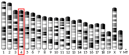
MUTYH has its locus on the short (p) arm of chromosome 1 (1p34.1), from base pair 45,464,007 to base pair 45,475,152 (45,794,835–45,806,142). The gene is composed of 16 exons and has a size of 546 amino acids[6] and is approximately 7.1kb.[7] The presence of disulfide crosslinking gives rise to a complex crystal structure of the MUTY-DNA.[8] The protein structure of the MUTYH gene has its N-terminal on the 5’ and the C-terminal on the 3’. Within the N-terminal. There is an helix-hairpin-helix and pseudo helix-hairpin-helix contained within the N-terminal, in addition to an iron cluster motif
Mechanism[]
Repair of oxidative DNA damage is the result of a collaborative effort of MUTYH, OGG1, and MTH1. MUTYH gene acts on the adenine base that have an A to 8-oxoG pairing while OGG1 (on chromosome 3 (3p26.2) part of the base excision repair pathway) detects and acts on 8-oxoG, thereby removing it.[9][10] The resultant effect of the action of the genes results in correction of transversion mutations made by the incorrect G:C, T:A pairing. TP53 transcriptionally regulates MUTYH and it can be surmised that it may potentially act as a regulator for p53.[11]
Expression[]
MUTYH is overexpressed in CD4-T cells, the prostate, the colon and the rectum. There is evidence of MUTYH expression in kidney, intestinal, nervous system and muscle tissues.[6]
Protein interactions[]
MUTYH has been shown to interact with Replication protein A1,[12] PCNA[12] and APEX1.[12]
The excision of the bases causes the formation of an apurinic/ apyrimidinic (AP site) gap. These gap sites are mutagenic in nature and require constant and immediate emendation and this is achieved by the active involvement of protein complexes that repair the AP gap site via short and long patch repair pathways. The short patch repair pathway employs POLB (DNA polymerase beta), APE1, XRCC1, PARP1 with the addition of either the LIG1 or LIG3 genes. When an insertion of one nucleotide occurs, the enzyme AP endonuclease (APEX/APE1) cuts out the mismatched base pairs at the AP site and this causes the evolvement of 5’dRP (5’ deoxyribose phosphate), a terminal blocking group, and 3’-OH ( 3’ hydroxyl end). POLB is required to remove the 5’dRP, and it does this by enzymatic activity, namely polymerase and dRP lyase. DNA ligase is used to seal the fragments after dRP excision causes the formation of 5’PO4 that is necessary to form the phosphodiester bonds of DNA. The purpose of PARP1 and XRCC1 in the single strand break repair pathway, is to stabilize the strands of DNA while they undergo repair, synthesis, gap-filling and ligation. PARP1 acts as a recruit agent for XRCC1. The nick sealing of the strands is accomplished by the formation of LIG1 (DNA ligase 1) and/or LIG3/ XRCCI complex that attach to processed end of the corrected strands and reinstate the original conformation of the strand. Long patch repair comes into play when more nucleotides are involved, ranging from 2 to 12. It is hypothesized that Polymerase