Median cubital vein
| Median cubital vein | |
|---|---|
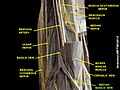
 Superficial veins of the upper limb. The median cubital vein is labelled (in Latin) - Vena mediana cubiti. | |
| Details | |
| Source | cephalic vein |
| Drains to | basilic vein |
| Identifiers | |
| Latin | vena mediana cubiti |
| TA98 | A12.3.08.019 |
| TA2 | 4980 |
| FMA | 22963 |
| Anatomical terminology | |
In human anatomy, the median cubital vein (or median basilic vein) is a superficial vein of the upper limb.[1] It lies in the cubital fossa superficial to the bicipital aponeurosis. It connects the cephalic vein and the basilic vein. It becomes prominent when pressure is applied. It is routinely used for venipuncture (taking blood) and as a site for an intravenous cannula. This is due to its particularly wide lumen, and its tendency to remain stationary upon needle insertion.
Structure[]
The median cubital vein is a superficial vein of the upper limb.[1] It lies in the cubital fossa superficial to the bicipital aponeurosis. It connects the cephalic vein and the basilic vein.[2] It becomes prominent when pressure is applied to upstream veins, as venous blood builds up.[3]
Variations[]
The median cubital vein shows a wide range of variations. More commonly, the vein forms an H-pattern with the cephalic and basilic veins making up the sides.[4] Other forms include an M-pattern, where the vein branches to the cephalic and basilic veins.[4]
Clinical significance[]
The median cubital vein is routinely used for venipuncture (taking blood) and as a site for an intravenous cannula.[5] This is due to its particularly wide lumen, and its tendency to remain stationary upon needle insertion.[5] It becomes prominent when pressure is applied upstream, which makes needle insertion easier.[3] Such pressure is created using a tourniquet.[3]
Additional images[]

The most frequent variations of the veins of the forearm (schematic).[4]
The median cubital vein, labelled as the median basilic vein.


See also[]
References[]
- ^ a b Standring, Susan (2016). Gray's anatomy: the anatomical basis of clinical practice (41 ed.). Elsevier Limited. pp. 837–861. ISBN 978-0-7020-5230-9.
- ^ Hunter, James P.; Barlow, Adam D.; Nicholson, Michael L. (2014). "5 - Access for Renal Replacement Therapy". Kidney Transplantation–Principles and Practice (7th ed.). Saunders. pp. 72–90. doi:10.1016/B978-1-4557-4096-3.00005-2. ISBN 978-1-4557-4096-3.
- ^ a b c Sadrzadeh, Hossein; Baskin, Leland; Kline, Gregory (2017). "1 - Variables affecting endocrine tests results, errors prevention and mitigation". Endocrine Biomarkers - Clinicians and Clinical Chemists in Partnership. Elsevier. pp. 1–40. doi:10.1016/B978-0-12-803412-5.00001-X. ISBN 978-0-12-803412-5.
- ^ a b c Pires, L.; Ráfare, A. L.; Peixoto, B. U.; Pereira, T. O. J. S.; Pinheiro, D. M. M.; Siqueira, M. E. B.; Vaqueiro, R. D.; de Paula, R. C.; Babinski, M. A.; Chagas, C. A. A. (2018-06-01). "The venous patterns of the cubital fossa in subjects from Brazil". Morphologie. 102 (337): 78–82. doi:10.1016/j.morpho.2018.02.001. ISSN 1286-0115. PMID 29625795.
- ^ a b Lew, K. (2012-01-01), Pawliszyn, Janusz (ed.), "3.05 - Blood Sample Collection and Handling", Comprehensive Sampling and Sample Preparation, Oxford: Academic Press, pp. 95–121, doi:10.1016/b978-0-12-381373-2.00068-5, ISBN 978-0-12-381374-9, retrieved 2020-11-17
External links[]
- Anatomy photo:07:st-0703 at the SUNY Downstate Medical Center
- Radiology image: UpperLimb:18VenoFo from Radiology Atlas at SUNY Downstate Medical Center (need to enable Java)
| Authority control: Scientific databases |
|---|
- Veins of the upper limb