Posterior longitudinal ligament
The posterior longitudinal ligament is a ligament connecting the posterior surfaces of the vertebral bodies of all of the vertebrae. It weakly prevents hyperflexion of the vertebral column. It also prevents posterior spinal disc herniation, although problems with the ligament can cause it.
Structure[]
| Posterior longitudinal ligament | |
|---|---|
 Posterior longitudinal ligament, in the thoracic region. (Posterior longitudinal ligament runs vertically at center.) | |
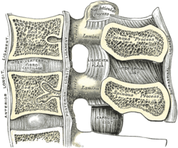 Median sagittal section of two lumbar vertebrae and their ligaments. (Posterior longitudinal ligament runs vertically at center left.) | |
| Details | |
| System | skeletal |
| Identifiers | |
| Latin | ligamentum longitudinale posterius |
| TA98 | A03.2.01.008 |
| TA2 | 1680 |
| FMA | 31894 |
| Anatomical terminology | |
The posterior longitudinal ligament is situated within the vertebral canal. It extends along the posterior surfaces of the bodies of the vertebrae, from the body of the axis to the sacrum and possibly the coccyx.[1] It is continuous with the tectorial membrane of atlanto-axial joint.[1] The ligament is thicker in the thoracic than in the cervical and lumbar regions. In the thoracic and lumbar regions, it presents a series of dentations with intervening concave margins.
The posterior longitudinal ligament is narrow at the vertebral bodies, where it covers the basivertebral veins, and widens at the intervertebral disc space. It is generally quite wide and thin.[1]
This ligament is composed of smooth, shining, longitudinal fibers, denser and more compact than those of the anterior ligament, and consists of superficial layers occupying the interval between three or four vertebræ, and deeper layers which extend between adjacent vertebrae.[2] Deep fibres run between each vertebral body.[1] Superficial fibres run between multiple vertebrae.[1]
Function[]
The posterior longitudinal ligament weakly prevents hyperflexion of the vertebral column.[3] It also limits spinal disc herniation, although it is much narrower than the anterior longitudinal ligament.[3]
Clinical significance[]
The posterior longitudinal ligament is much narrower than the anterior longitudinal ligament.[3] Because of this, spinal disc herniations usually occur in a posterolateral direction.[3]
The posterior longitudinal ligament contains a higher density of nociceptors than many ligaments, so can cause back pain.[1] It may ossify, particularly around cervical vertebrae.[1]
The posterior longitudinal ligament has a high density of vasomotor fibres, allowing for increased blood flow to respond to damage to the ligament.[1]
See also[]
References[]
- ^ a b c d e f g h Cramer, Gregory D. (2014). "5 - The Cervical Region". Clinical anatomy of the spine, spinal cord, and ANS (3rd ed.). St. Louis: Mosby, Elsevier Health Sciences. pp. 135–209. doi:10.1016/B978-0-323-07954-9.00005-0. ISBN 978-0-323-07954-9. OCLC 830314791.
- ^
 This article incorporates text in the public domain from page 288 of the 20th edition of Gray's Anatomy (1918)
- ^ a b c d Moore, K.; Dalley, A.; Agur, A. (2018). Clinically Oriented Anatomy (8th ed.). pp. 98–108. ISBN 9781496347213.
Additional images[]
F: Posterior longitudinal ligament

Membrana tectoria, transverse, and alar ligaments.


External links[]
- Atlas image: back_bone25 at the University of Michigan Health System - "Vertebral Column, Dissection, Anterior & Posterior Views"
- lesson7 at The Anatomy Lesson by Wesley Norman (Georgetown University) - "Lateral Pharyngeal Region"
Authority control | |
|---|---|
| Scientific databases | |
| Other | |
This ligament-related article is a stub. You can help Wikipedia by . |
- Wikipedia articles incorporating text from the 20th edition of Gray's Anatomy (1918)
- Ligaments of the torso
- Bones of the vertebral column
- Ligaments of the head and neck
- Ligament stubs