Superior parietal lobule
| Superior parietal lobule | |
|---|---|
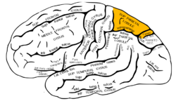 Lateral surface of left cerebral hemisphere, viewed from the side. (Superior parietal lobule is shown in orange.) | |
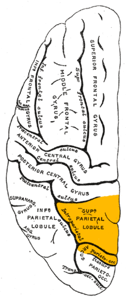 Lateral surface of left cerebral hemisphere, viewed from above. (Superior parietal lobule is shown in orange.) | |
| Details | |
| Identifiers | |
| Latin | lobulus parietalis superior |
| NeuroNames | 106 |
| TA98 | A14.1.09.130 |
| TA2 | 5476 |
| FMA | 61899 |
| Anatomical terms of neuroanatomy | |
The superior parietal lobule is bounded in front by the upper part of the postcentral sulcus, but is usually connected with the postcentral gyrus above the end of the sulcus. The superior parietal lobule contains Brodmann's areas 5 and 7.
Behind it is the lateral part of the parietooccipital fissure, around the end of which it is joined to the occipital lobe by a curved gyrus, the . Below, it is separated from the inferior parietal lobule by the horizontal portion of the intraparietal sulcus.
The superior parietal lobule is involved with spatial orientation,[1] and receives a great deal of visual input as well as sensory input from one's hand.[2] It is also involved with other functions of the parietal lobe in general.
There are major white matter pathway connections with the superior parietal lobule such as the Cingulum, , superior parietal lobule connections of the Medial longitudinal fasciculus and other newly described superior parietal white matter connections.[3][4]
Damage to the superior parietal lobule can cause contralateral astereognosis and hemispatial neglect. It is also associated with deficits on tests involving the manipulation and rearrangement of information in working memory, but not on working memory tests requiring only rehearsal and retrieval processes.[5]
Additional images[]
Position of superior parietal lobule (shown in red).

Superior parietal lobule (shown in red).

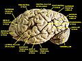
Superior parietal lobule is labeled at upper left.
Drawing to illustrate the relations of the brain to the skull.

Lateral view of a human brain, main gyri labeled.
Cerebrum. Lateral view. Deep dissection.

Cerebrum. Lateral view. Deep dissection.

Cerebrum. Lateral view. Deep dissection.

Superior parietal lobule highlighted in green on coronal T1 MRI images

Superior parietal lobule highlighted in green on sagittal T1 MRI images

Superior parietal lobule highlighted in green on transversal T1 MRI images


References[]
![]() This article incorporates text in the public domain from page 823 of the 20th edition of Gray's Anatomy (1918)
This article incorporates text in the public domain from page 823 of the 20th edition of Gray's Anatomy (1918)
- ^ Sylvius entry on "superior parietal lobe" Archived June 26, 2009, at the Wayback Machine
- ^ Brainmind.com "Parietal Area 5"
- ^ Kamali A, Sair HI, Radmanesh A, Hasan KM. (2014). Decoding the superior parietal lobule connections of the superior longitudinal fasciculus/arcuate fasciculus in the human brain. Neuroscience. 277:577-83. doi: 10.1016/j.neuroscience. PMID 25086308
- ^ Kamali, A; Flanders, AE; Brody, J; Hunter, JV; Hasan, KM (2014). Tracing superior longitudinal fasciculus connectivity in the human brain using high resolution diffusion tensor tractography. Brain Struct Funct. 219: 269–81. doi:10.1007/s00429-012-0498-y. PMID 23288254.
- ^ Koenigs, M., Barbey, A. K., Postle, B. R., & Grafman, J. (2009). Superior parietal cortex is critical for the manipulation of information in working memory. The Journal of Neuroscience, 29(47), 14980-14986.
| Wikimedia Commons has media related to Superior parietal lobule. |
| Authority control: Scientific databases |
|---|
This neuroanatomy article is a stub. You can help Wikipedia by . |
- Wikipedia articles incorporating text from the 20th edition of Gray's Anatomy (1918)
- Parietal lobe
- Gyri
- Neuroanatomy stubs