Arytenoid muscle
| Arytenoid muscle | |
|---|---|
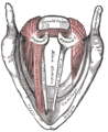
 Muscles of larynx. Posterior view. Oblique arytenoid: The "X" in the center. Transverse arytenoid: Bands underneath the "X". Aryepiglotticus: Wraps around back. | |
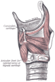
 Sagittal section of the larynx and upper part of the trachea. (Arytenoideus visible at center right.) | |
| Details | |
| Origin | Arytenoid cartilage on one side |
| Insertion | Arytenoid cartilage on opposite side |
| Artery | superior laryngeal artery |
| Nerve | recurrent laryngeal branch of the vagus |
| Actions | approximate the arytenoid cartilages (close rima glottis) |
| Identifiers | |
| Latin | Musculus arytaenoideus |
| TA98 | A06.2.08.012 A06.2.08.010 |
| TA2 | 2203, 2205 |
| FMA | 46582 |
| Anatomical terms of muscle | |
The arytenoid muscle /ærɪˈtiːnɔɪd/ is a single muscle of the larynx. It passes from one arytenoid cartilage to the opposite arytenoid cartilage. It has oblique and transverse fibres. It is supplied by the recurrent laryngeal nerve. It approximates the arytenoid cartilages. Continuous electromyography may be used during neck surgeries such as thyroidectomy.
Structure[]
The arytenoid muscle fills the posterior concave surface of the arytenoid cartilage. It arises from the posterior surface and lateral border of one arytenoid cartilage. It is inserted into the corresponding parts of the opposite arytenoid cartilage.
It consists of oblique and transverse fibres.
Nerve supply[]
The arytenoid muscle is supplied by the recurrent laryngeal nerve, a branch of the vagus nerve (CN X).[1] This is a bilateral supply.[2]
Function[]
The arytenoid muscle approximates the arytenoid cartilages. This closes the aperture of the glottis, especially at its back part to eliminate the posterior commissure of the vocal cords.
Clinical significance[]
Electromyography[]
Function of the arytenoid muscle is a good method to determine function of the recurrent laryngeal nerve.[1] Continuous electromyography of the arytenoid muscle can provide confidence to surgeons that the recurrent laryngeal nerve is not damaged during neck surgeries, such as thyroidectomy.[1]
Other animals[]
The arytenoid muscle is found in many animals, including dogs.[2]
Additional images[]

The cartilages of the larynx. Posterior view.
Muscles of larynx. Side view. Right lamina of thyroid cartilage removed.

Aryepiglotic muscle
Muscles of the larynx, seen from above.

Dissection of the muscles of the palate from behind.


References[]
 This article incorporates text in the public domain from page 1082 of the 20th edition of Gray's Anatomy (1918)
- ^ a b c Li, Peng; Liang, Qing-Zhuang; Wang, Dong-Lai; Han, Bin; Yi, Xin; Wei, Wei (October 2019). "Modified arytenoid muscle electrode recording method for neuromonitoring during thyroidectomy". Gland Surgery. 8 (5): 469–476. doi:10.21037/gs.2019.08.07. ISSN 2227-684X. PMC 6842767. PMID 31741877.
- ^ a b 隆一, 相原 (1991). "イヌ披裂筋の構造と運動神経支配に関する研究". 日本耳鼻咽喉科学会会報. 94 (6): 805–816. doi:10.3950/jibiinkoka.94.805. PMID 1715914.
Authority control | |
|---|---|
| Scientific databases | |
| Other | |
- Wikipedia articles incorporating text from the 20th edition of Gray's Anatomy (1918)
- Muscles of the head and neck