Sternothyroid muscle
| Sternothyroid muscle | |
|---|---|
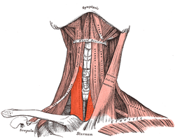 Sternothyroid visible center left | |
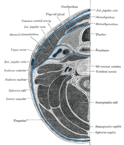 Section of the neck at about the level of the sixth cervical vertebra. Showing the arrangement of the fascia coli. (Sternothyroideus labeled at right, third from top.) | |
| Details | |
| Origin | Manubrium |
| Insertion | Thyroid cartilage |
| Artery | Superior thyroid artery |
| Nerve | Ansa cervicalis |
| Actions | Depresses thyroid cartilage |
| Identifiers | |
| Latin | Musculus sternothyroideus |
| TA98 | A04.2.04.006 |
| TA2 | 2173 |
| FMA | 13343 |
| Anatomical terms of muscle | |
The sternothyroid muscle, or sternothyroideus, is an infrahyoid muscle in the neck.[1] It acts to depress the hyoid bone.[2] It is below the sternohyoid muscle. It is shorter and wider than the sternohyoid.
Structure[]
The sternothyroid arises from the posterior surface of the manubrium of the sternum, below the origin of the sternohyoid.[3] It also arises from the edge of the cartilage of the first rib. It is inserted into the oblique line on the lamina of the thyroid cartilage. It is in close contact with its fellow at the lower part of the neck, but diverges somewhat as it ascends. It is occasionally traversed by a transverse or oblique tendinous inscription.
Innervation[]
The sternothyroid muscle is innervated by the ansa cervicalis.[4][5]
Variations[]
Doubling; absence; accessory slips to the thyrohyoid, inferior pharyngeal constrictor, or to the carotid sheath.
Function[]
The sternothyroid muscle depresses the hyoid bone, along with the other infrahyoid muscle.[2]
Clinical significance[]
The upward extension of a thyroid swelling (goitre) is prevented by the attachment of the sternothyroid to the thyroid cartilage. A goitre can therefore only grow to the front, back or middle but no higher.
Additional images[]

Superficial dissection of the right side of the neck, showing the carotid and subclavian arteries.
The fascia and middle thyroid veins.

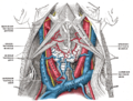
Hypoglossal nerve, cervical plexus, and their branches.

Side view of the larynx, showing muscular attachments.

Sternothyroid muscle


References[]
![]() This article incorporates text in the public domain from page 393 of the 20th edition of Gray's Anatomy (1918)
This article incorporates text in the public domain from page 393 of the 20th edition of Gray's Anatomy (1918)
- ^ Chokroverty, Sudhansu (2009-01-01), Chokroverty, Sudhansu (ed.), "Chapter 7 - Physiologic Changes in Sleep", Sleep Disorders Medicine (Third Edition), Philadelphia: W.B. Saunders, pp. 80–104, doi:10.1016/b978-0-7506-7584-0.00007-0, ISBN 978-0-7506-7584-0, retrieved 2020-11-25
- ^ a b Derksen, Frederik J. (2006-01-01), Auer, Jörg A.; Stick, John A. (eds.), "Chapter 40 - Overview of Upper Airway Function", Equine Surgery (Third Edition), Saint Louis: W.B. Saunders, pp. 516–522, doi:10.1016/b1-41-600123-9/50042-5, ISBN 978-1-4160-0123-2, retrieved 2020-11-25
- ^ Fessler, Richard G.; Kim, Daniel H. (2012-01-01), Quiñones-Hinojosa, Alfredo (ed.), "Chapter 191 - Surgical Approaches to the Cervicothoracic Junction", Schmidek and Sweet Operative Neurosurgical Techniques (Sixth Edition), Philadelphia: W.B. Saunders, pp. 2177–2191, doi:10.1016/b978-1-4160-6839-6.10191-1, ISBN 978-1-4160-6839-6, retrieved 2020-11-25
- ^ McHanwell, Steve; Watson, Charles (2009-01-01), Watson, Charles; Paxinos, George; Kayalioglu, Gulgun (eds.), "Chapter 7 - Localization of Motoneurons in the Spinal Cord", The Spinal Cord, San Diego: Academic Press, pp. 94–114, doi:10.1016/b978-0-12-374247-6.50011-0, ISBN 978-0-12-374247-6, retrieved 2020-11-25
- ^ Cesmebasi, Alper (2015-01-01), Tubbs, R. Shane; Rizk, Elias; Shoja, Mohammadali M.; Loukas, Marios (eds.), "Chapter 31 - Anatomy of the Cervical Plexus and Its Branches", Nerves and Nerve Injuries, San Diego: Academic Press, pp. 441–449, doi:10.1016/b978-0-12-410390-0.00032-9, ISBN 978-0-12-410390-0, retrieved 2020-11-25
External links[]
- Photo of model at Waynesburg College musclehead/sternothyroid
- Anatomy photo:25:03-0105 at the SUNY Downstate Medical Center - "The Muscular triangle"
- PTCentral
| Authority control: Scientific databases |
|---|
- Wikipedia articles incorporating text from the 20th edition of Gray's Anatomy (1918)
- Muscles of the head and neck