Basilar artery
| Basilar artery | |
|---|---|
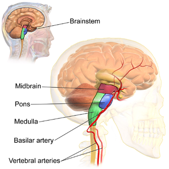 The basilar artery lies at the front of the brainstem in the midline and is formed from the union of the two vertebral arteries. | |
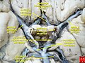
 Diagram of the arterial circulation at the base of the brain (inferior view). The basilar artery terminates by splitting into the left and right posterior cerebral arteries. | |
| Details | |
| Source | Vertebral arteries |
| Branches | Pontine arteries, anterior inferior cerebellar (AICA) and superior cerebellar arteries, and terminal posterior cerebral arteries. |
| Supplies | Pons, and superior and inferior aspects of the cerebellum. |
| Identifiers | |
| Latin | Arteria basilaris |
| MeSH | D001488 |
| TA98 | A12.2.07.081 |
| TA2 | 4548 |
| FMA | 50542 |
| Anatomical terminology | |
The basilar artery (/ˈbæz.ɪ.lər/)[1][2] is one of the arteries that supplies the brain with oxygen-rich blood.
The two vertebral arteries and the basilar artery are known as the vertebral basilar system, which supplies blood to the posterior part of the circle of Willis and joins with blood supplied to the anterior part of the circle of Willis from the internal carotid arteries.[3][4][5]
Structure[]
The basilar artery arises from the union of the two vertebral arteries at the junction between the medulla oblongata and the pons between the abducens nerves (CN VI).[6]
It ascends superiorly in the basilar sulcus of the ventral pons and divides at the junction of the midbrain and pons into the posterior cerebral arteries.
Its branches from caudal to rostral include:
- anterior inferior cerebellar artery
- labyrinthine artery (<15% of people, usually branches from the anterior inferior cerebellar artery)
- pontine arteries
- superior cerebellar artery
Clinical relevance[]
A basilar artery stroke classically leads to locked-in syndrome.
Additional images[]

The internal carotid and vertebral arteries (Right side view)
Basilar artery
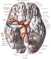
The arteries of the base of the brain. Basilar artery labeled below center. The temporal pole of the cerebrum and the cerebellar hemisphere have been removed on the right side. Inferior aspect (viewed from below).


References[]
- ^ "BASILAR | meaning in the Cambridge English Dictionary". dictionary.cambridge.org.
- ^ "basilar - WordReference.com Dictionary of English". www.wordreference.com.
- ^ Jones, Jeremy. "Basilar artery | Radiology Reference Article | Radiopaedia.org". Radiopaedia.
- ^ Purves, Dale (2012). Neuroscience (5th ed.). Sunderland, Mass. pp. 737–738. ISBN 9780878936953.
- ^ Carpenter, Malcolm B. (1985). Core text of neuroanatomy (3rd ed.). Baltimore: Williams & Wilkins. pp. 406–410. ISBN 0683014552.
- ^ Byrne, James (2012). "Chapter 2. Cranial arterial anatomy". Tutorials in endovascular neurosurgery and interventional neuroradiology. Berlin: Springer. pp. 37–38.
External links[]
| Wikimedia Commons has media related to Arteria basilaris. |
- Basilar Artery at neuroangio.org
- Anatomy photo:28:09-0204 at the SUNY Downstate Medical Center - "Cranial Fossae: Arteries, Inferior Surface of the Brain"
- Blood supply at neuropat.dote.hu
- "Anatomy diagram: 13048.000-1". Roche Lexicon - illustrated navigator. Elsevier. Archived from the original on 2014-01-01.
- "Anatomy diagram: 13048.000-3". Roche Lexicon - illustrated navigator. Elsevier. Archived from the original on 2014-01-01.
| Authority control: Scientific databases |
|---|
- Arteries of the head and neck