Capsule of lens
This article needs additional citations for verification. (July 2020) |
| Capsule of lens | |
|---|---|
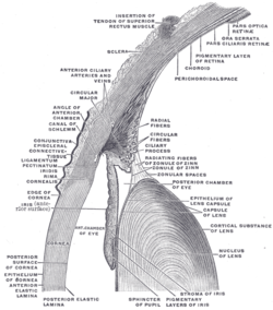
 The upper half of a sagittal section through the front of the eyeball. (Capsule of lens labeled at center right.) | |
| Details | |
| Identifiers | |
| Latin | capsula lentis |
| MeSH | D007903 |
| TA98 | A15.2.05.007 |
| FMA | 58881 |
| Anatomical terminology | |
The lens capsule is a component of the globe of the eye. It is a clear, membrane-like structure composed of collagen IV and laminin that is quite elastic, a quality that keeps it under constant tension. As a result, the lens naturally tends towards a rounder or more globular configuration, a shape it must assume for the eye to focus at a near distance. Lens capsule is the thickest basement membrane in the body.[1]
Normally, the lens capsule serves as a diffusion barrier. It is permeable to low molecular weight compounds but restricts the movement of large colloidal particles.[2]
Anatomy[]
The lens capsule is a transparent membrane that surrounds the entire lens. The capsule is thinnest at the posterior pole with approximate thickness of 3.5μm. Average thickness at the equator is 7μm. Anterior pole thickness increases with age from 11-15μm. The thickest portion of is annular region surrounding the anterior pole. This will also increases with age (from 13.5-16μm).[3]
Even though the capsule is a highly elastic structure, it contains no elastic fibers. Elasticity is because of the thick lamellar arrangement of the collagen fibers.[3]
Embryology[]
The lens vesicle is developed from surface ectoderm. It will separate from surface ectoderm at approximately day 33. Lens capsule developed from basal lamina of lens vesicle will cover early lens fibers. Capsule is evident at 5 weeks of gestation.[3]
Vascular lens capsule[]
During fetal development vascular lens capsule (tunica vasculosa lentis) develop from the mesenchyme that surrounds the lens. It receives arterial blood supply from the hyaloid artery.[2] This blood supply slowly regress and vascular capsule disappear before birth. The disappearance of the anterior vascular capsule of the lens is useful in estimating the gestational age.[4]
Function[]
The capsule helps give the lens its spherical shape.[5]
Accommodation[]
Normally, when ciliary muscles are in relaxed state, the zonules will pull the capsule. Due to this zonular tension anterior lens surface becomes flat. When ciliary muscles contract, this zonular tension will reduce allowing lens to assume more spherical shape. This shape change increase the whole power of the eye, and people will be able to see near clearly. The process of changing lens power to see near clearly is known as accommodation.
Lens protection[]
Early embryologic development of lens capsule give lens materiel an immune privilege. It will also help protecting lens from virus and bacteria.[3]
Clinical significance[]
In intra-capsular cataract extraction (ICCE), whole lens including capsule is removed. During more common extra capsular cataract surgery procedures like micro inscision cataract surgery, phacoemulsification etc., clouded lens is removed through opening made in anterior lens capsule. The intraocular lens is then inserted into the lens capsule. The best place for intraocular lens implantation is within the capsular bag.[6]
Posterior capsular opacification and posterior capsule rupture are common complications of cataract surgery.[7]
See also[]
References[]
- ^ Yanoff, Myron. (2009). "Lens". Ocular pathology. Sassani, Joseph W. (6th ed.). Edinburgh: Mosby/Elsevier. ISBN 978-0-323-04232-1. OCLC 294998596.
- ^ a b Snell, Richard S. (2012). "Development of the Eye and the Ocular Appendages". Clinical anatomy of the eye. Lemp, Michael A. (2nd ed.). Malden, MA, USA: Blackwell Science. ISBN 978-0-632-04344-6. OCLC 37580703.
- ^ a b c d Clinical anatomy and physiology of the visual system (3rd ed.). Elsevier/Butterworth-Heinemann. 2012. ISBN 978-1-4377-1926-0.
- ^ Hittner, H. M.; Hirsch, N. J.; Rudolph, A. J. (September 1977). "Assessment of gestational age by examination of the anterior vascular capsule of the lens". The Journal of Pediatrics. 91 (3): 455–458. doi:10.1016/s0022-3476(77)81324-3. ISSN 0022-3476. PMID 894419.
- ^ "Lens Capsule". American Academy of Ophthalmology. 1 October 2019.
- ^ Mehta, Rajvi; Aref, Ahmad A (27 November 2019). "Intraocular Lens Implantation In The Ciliary Sulcus: Challenges And Risks". Clinical Ophthalmology (Auckland, N.Z.). 13: 2317–2323. doi:10.2147/OPTH.S205148. ISSN 1177-5467. PMC 6885568. PMID 31819356.
- ^ John F, Salmon (13 December 2019). Kanski's clinical ophthalmology : a systematic approach (9th ed.). Elsevier. ISBN 978-0-7020-7711-1.
- Human eye anatomy

