Germinal center
This article needs additional citations for verification. (May 2016) |
| Germinal center | |
|---|---|
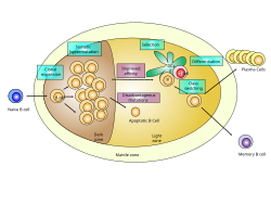 Germinal center of a lymph node showing proliferation and development stages of a B cell . | |
| Identifiers | |
| MeSH | D018858 |
| Anatomical terminology | |
Germinal centers or germinal centres (GCs) are transiently formed structures within B cell zone (follicles) in secondary lymphoid organs – lymph nodes, ileal Peyer's patches, and the spleen[1] – where mature B cells are activated, proliferate, differentiate, and mutate their antibody genes (through somatic hypermutation aimed at achieving higher affinity) during a normal immune response; most of the germinal center B cells (BGC) are removed by tingible body macrophages.[2] The B cells develop dynamically after the activation of follicular B cells by T-dependent antigen.
As they undergo rapid and mutative cellular division, B cells of the germinal center's dark zone are known as centroblasts. Once these B cells have stopped proliferating and moved to the light zone, they are known as centrocytes, and are subjected to selection by follicular helper T (TFH) cells in the presence of follicular dendritic cells (FDCs). Germinal centers are an important part of the B cell humoral immune response, acting as central factories for the generation of affinity matured B cells specialized in producing improved antibodies that effectively recognize antigen (e.g. infectious agents), and for the production of long-lived plasma cells and durable memory B cells.
Process[]

- Centrocytes are small to medium size with angulated, elongated, cleaved, or twisted nuclei.
- Centroblasts are larger cells containing vesicular nuclei with one to three basophilic nucleoli apposing the nuclear membrane.
- Follicular dendritic cells have round nuclei, centrally located nucleoli, bland and dispersed chromatin, and flattening of adjacent nuclear membrane.
- Within lymph nodes, mature peripheral B cells known as follicular (Fo) B cells acquire antigen from FDCs and in turn present it to cognate CD4+ TFH cells at the border that demarcates the interfollicular T cell area and B cell zone (also known as lymphoid follicles).
- After several rounds of cellular division, the B cells go through somatic hypermutation, a process by which they mutate their antibody-encoding DNA and thus generate a diversity of clones in the germinal center. This involves pseudo-random substitutions biased towards regions encoding the antigen recognition surface of the antibodies the B cells produce. This phenomenon underscores the process of affinity maturation, whereby greater affinity antibodies are produced and selected for after antigen recognition.
- Upon receiving an unidentified stimulus, the maturing B cells (centroblasts) migrate from the dark zone to the light zone and start to express their edited BCRs on the cell surface and at this stage are referred to as centrocytes. The centrocytes are in a state of activated apoptosis and compete for survival signals derived from FDCs and TFH cells. This rescue process, known as germinal center selection, is believed to be dependent on the affinity of their surface antibody to the antigen. Such that, a B cell that has successfully gained mutations that confer a higher affinity surface antibody towards antigen gains a survival advantage over lower affinity B cell clones and those that have gained deleterious mutations. Cyclic re-entry into the dark zone once again as centroblasts allows a chance for otherwise non-selected B cell mutants to gain more mutations in order to improve affinity towards antigen. Interactions with T cells are also believed to prevent the generation of autoreactive germinal center B cells.[3]
- At some unclear stage of their centroblast-centrocyte cycling, maturing B cells receive a final differentiation signal to exit the germinal center as an antibody producing plasma cell which are cells that secrete large quantities of antibody or a memory B cell that can be reactivated in subsequent contacts with the same antigen. Selected B cells may also restart the whole cycle of mutative centroblast division and centrocyte selection. In this way the adaptive immune system, in part through these germinal center reactions, can gradually better recognize antigens over time.
Morphology at different stages[]
The morphology of GCs is very specific and shows properties which are characteristic for different stages of the reaction.
- In an early state of the reaction a network of FDCs is fully filled with proliferating B cells.
- Later at day 4 of the reaction, GCs show a separation of two zones, the dark and the light zone.[4] The former still contains dominantly proliferating and mutating B cells while the latter one is the area of B cell selection.
- These zones dissolve after 10 days of GC development which ends after about 3 weeks.
Medical relevance[]
As germinal centers are important structures of the adaptive immune system, their deregulation is implied in many immune diseases, for example rheumatoid arthritis, immunodeficiency and many lymphomas like DLBCL and Burkitt's lymphoma.
See also[]
- Lymphatic system
- Tingible body
- Mantle zone
References[]
- ^ (1 January 2007). "The Biology of the Germinal Center". Hematology. 2007: 210–215. doi:10.1182/asheducation-2007.1.210. PMID 18024632.
- ^ Aguzzi A, Kranich J, Krautler NJ (2014). "Follicular dendritic cells: origin, phenotype, and function in health and disease". Trends in Immunology. 35 (3): 105–113. doi:10.1016/j.it.2013.11.001. PMID 24315719.
- ^ Thorbecke GJ, Amin AR, Tsiagbe VK (1994). "Biology of germinal centres in lymphoid tissue". FASEB Journal. 8 (11): 832–840. doi:10.1096/fasebj.8.11.8070632. PMID 8070632. S2CID 83999556.
- ^ Meyer-Hermann ME (2002). "A Mathematical Model for the Germinal Centre Morphology and Affinity Maturation". J. Theor. Biol. 216 (3): 273–300. arXiv:physics/0203021. Bibcode:2002JThBi.216..273M. doi:10.1006/jtbi.2002.2550. PMID 12183119. S2CID 2141888.
External links[]
- UIUC Histology Subject 563
- Histology image: 07103loa – Histology Learning System at Boston University - "Lymphoid Tissues and Organs: lymph node, germinal centre"
- Hyperlinked Human Histology
- MedEd at Loyola Histo/practical/lymph/hp12-42.html
- Lymphatic system
- Lymphatic tissue