Hemangiopericytoma
| Hemangiopericytoma | |
|---|---|
| Other names | HPC |
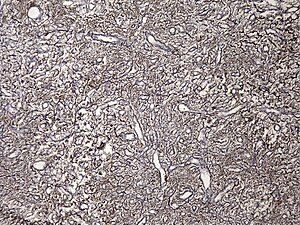 | |
| Haemangiopericytoma, Gomori methenamine silver stain | |
| Specialty | Neuro-oncology |
| Symptoms | Painless mass[1] |
| Usual onset | 45 years of age (median)[1] |
A hemangiopericytoma is a type of soft-tissue sarcoma that originates in the pericytes in the walls of capillaries. When inside the nervous system, although not strictly a meningioma tumor, it is a meningeal tumor with a special aggressive behavior. It was first characterized in 1942.[2]
Histopathology[]
Hemangiopericytomas are tumors that are derived from specialized spindle shaped cells called pericytes, which line capillaries.[3]
Diagnosis[]
Hemangiopericytoma located in the cerebral cavity is an aggressive tumor of the mesenchyme with oval nuclei with scant cytoplasm. "There is dense intercellular reticulin staining. Tumor cells can be fibroblastic, myxoid, or pericytic. These tumors, in contrast to meningiomas, do not stain with epithelial membrane antigen. They have a grade 2 or 3 biological behavior, and need to be distinguished from benign meningiomas because of their high rate of recurrence (68.2%) and metastases (Maier et al. 1992;[4] Kleihues et al. 1993 [5])."[6]
Treatment[]
Depending on the grade of the sarcoma, it is treated with surgery,[7] chemotherapy, and/or radiotherapy.
Epidemiology[]
In one series, the median age of affected individuals was 45 years, with a 10 year survival rate of 70 percent.[1]
See also[]
- Infantile hemangiopericytoma
- List of cutaneous conditions
References[]
- ^ a b c Enzinger, FM; Smith, BH (January 1976). "Hemangiopericytoma. An analysis of 106 cases". Human Pathology. 7 (1): 61–82. doi:10.1016/s0046-8177(76)80006-8. PMID 1244311.
- ^ Stout AP, Murray MR (1942). "Hemangiopericytoma: a vascular tumor featuring Zimmermann's pericytes". Ann Surg. 116 (1): 26–33. doi:10.1097/00000658-194207000-00004. PMC 1543753. PMID 17858068.
- ^ Gerner, RE; Moore, GE; Pickren, JW (February 1974). "Hemangiopericytoma". Annals of Surgery. 179 (2): 128–32. doi:10.1097/00000658-197402000-00002. PMC 1355764. PMID 4359454. S2CID 220588367.
- ^ Maier H, Ofner D, Hittmair A, Kitz K, Budka H (1992). "Classic, atypical, and anaplastic meningioma: three histopathological subtypes of clinical relevance". Journal of Neurosurgery. 77 (4): 616–23. doi:10.3171/jns.1992.77.4.0616. PMID 1527622.
- ^ Kleihues P, Burger PC, Scheithauer BW (1993). "Histological typing of tumours of the central nervous system". World Health Organization. Berlin : Springer-Verlag (2nd ed.) (30).
- ^ Sherman Sojka W MD; Raizer J MD; Dropcho E.J.MD. "Meningiomas". Medmerits: 2. Archived from the original on 2012-03-31. Retrieved 2011-09-19.
- ^ Ozaki N, Mukohara N, Yoshida M, Shida T (April 2006). "Successful resection of giant hemangeopericytoma originating from the left atrium". Interact Cardiovasc Thorac Surg. 5 (2): 79–80. doi:10.1510/icvts.2005.124107. PMID 17670519.
Further reading[]
- Schiariti, M; Goetz, P; El-Maghraby, H; Tailor, J; Kitchen, N (Mar 2011). "Hemangiopericytoma: long-term outcome revisited. Clinical article". Journal of Neurosurgery. 114 (3): 747–55. doi:10.3171/2010.6.JNS091660. PMID 20672899.
External links[]
| Classification | |
|---|---|
| External resources |
- Dermal and subcutaneous growths
- Vascular neoplasia
- Nervous system neoplasia
- Brain tumor