Lipoblast

Histopathology of liposarcoma, H&E stain, with the main features:[1]
- Spindle cells with enlarged, hyperchromatic nuclei.
- Apparently univacuolated adipocytes (may look normal).
- Lipoblasts (multivacuolated), but neither necessary nor sufficient for diagnosis of liposarcoma.
A lipoblast is a precursor cell for an adipocyte.[2]
Alternate terms include adipoblast[3] and preadipocyte.[4]
Early stages are almost indistinguishable from fibroblasts.[5]
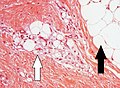
Lipoblasts (white arrow) and lipocytes (black arrow), in a case of lipoblastoma
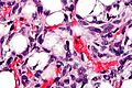
Micrograph showing a lipoblast (left-bottom of image) in a liposarcoma. H&E stain.


Liposarcoma[]
Lipoblasts are seen in liposarcoma[6] and characteristically have abundant multivacuolated clear cytoplasm and a dark staining (hyperchromatic), indented nucleus.
See also[]
- Adipogenesis
- Adipose differentiation-related protein
- Lipoblastoma
References[]
- ^ Michael R. Clay, M.D. "Liposarcoma". PathologyOutlines. Topic Completed: 1 November 2017. Minor changes: 11 May 2021
- ^ Barbara Young; Paul R. Wheater (2006). Wheater's functional histology: a text and colour atlas. Elsevier Health Sciences. pp. 74–. ISBN 978-0-443-06850-8. Retrieved 18 April 2010.
- ^ Dani C (1999). "Embryonic stem cell-derived adipogenesis". Cells Tissues Organs (Print). 165 (3–4): 173–80. doi:10.1159/000016697. PMID 10592389.
- ^ Coskun H, Summerfield TL, Kniss DA, Friedman A (April 2010). "Mathematical modeling of preadipocyte fate determination". J Theor Biol. 265 (1): 87–94. doi:10.1016/j.jtbi.2010.03.047. PMID 20385145.
- ^ Ray C. Henrikson; Gordon I. Kaye; Joseph E. Mazurkiewicz (31 July 1997). Histology. Lippincott Williams & Wilkins. pp. 118–. ISBN 978-0-683-06225-0. Retrieved 18 April 2010.
- ^ Lester J. Layfield (2002). Cytopathology of bone and soft tissue tumors. Oxford University Press US. pp. 83–. ISBN 978-0-19-513236-6. Retrieved 18 April 2010.
This article related to pathology is a stub. You can help Wikipedia by . |
Categories:
- Cell biology
- Pathology stubs