Posterior grey column
This article needs additional citations for verification. (September 2015) |
| Posterior grey column (Posterior horn of spinal cord) | |
|---|---|
 Cross section of the spinal cord. The posterior horn is the upper protrusion of grey matter, labeled with "2" | |
| Details | |
| Identifiers | |
| Latin | cornu posterius medullae spinalis |
| MeSH | D066148 |
| TA98 | A14.1.02.115 A14.1.02.023 A14.1.02.114 |
| TA2 | 6064 |
| FMA | 256530 |
| Anatomical terminology | |
The posterior grey column (posterior cornu, dorsal horn, spinal dorsal horn, posterior horn, sensory horn[1]) of the spinal cord is one of the three grey columns of the spinal cord. It receives several types of sensory information from the body, including fine touch, proprioception, and vibration. This information is sent from receptors of the skin, bones, and joints through sensory neurons whose cell bodies lie in the dorsal root ganglion.
Anatomy[]
The posterior grey column is subdivided into six layers termed Rexed laminae I-VI
- Marginal nucleus of spinal cord (lamina I)
- Substantia gelatinosa of Rolando (lamina II)
- Nucleus proprius (laminae III, IV)
- Spinal lamina V, the neck of the posterior horn[2]
- Spinal lamina VI, the base of the posterior horn.
The other four Rexed laminae are located in the other two grey columns in the spinal cord.
Additional images[]
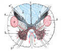
Section of the medulla oblongata through the lower part of the decussation of the pyramids

See also[]
- Posterior column-medial lemniscus pathway
- Posterior horn of lateral ventricles
- Anterior grey column
References[]
- ^ "Dorsal horn | anatomy". Encyclopedia Britannica. Retrieved 2021-05-04.
- ^ Woolsey, Robert M.; Vernon W. Lin; Cardenas, Diana D.; Cutter, Nancy C.; Frederick S. Frost; Margaret C. Hammond; Laurie B. Lindblom; Inder Perkash; Robert Waters (2002). Spinal Cord Medicine: Principles and Practice. Demos Medical Publishing. ISBN 1-888799-61-7.
| Authority control: Scientific databases |
|---|
This neuroanatomy article is a stub. You can help Wikipedia by . |
- Spinal cord
- Neuroanatomy
- Neuroanatomy stubs