Pulmonary artery
| Pulmonary artery | |
|---|---|
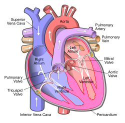 Anterior (frontal) view of the opened heart. White arrows indicate normal blood flow. (Pulmonary artery labeled at upper right.) | |
| Details | |
| Precursor | truncus arteriosus |
| System | Cardiovascular, Respiratory |
| Source | right ventricle |
| Identifiers | |
| Latin | arteria pulmonalis |
| MeSH | D011651 |
| TA98 | A12.2.01.101 A12.2.01.201 |
| TA2 | 4077, 4091 |
| FMA | 66326 |
| Anatomical terminology | |
A pulmonary artery is an artery in the pulmonary circulation that carries deoxygenated blood from the right side of the heart to the lungs. The largest pulmonary artery is the main pulmonary artery or pulmonary trunk from the heart, and the smallest ones are the arterioles, which lead to the capillaries that surround the pulmonary alveoli.
Structure[]
The pulmonary arteries are blood vessels that carry blood from the right side of the heart through to the capillaries of the lungs. The blood that is carried is, unlike other arteries, without oxygen ("deoxygenated"). The main pulmonary arteries emerge from the right side of the heart, and these split into smaller arteries that progressively divide and become smaller until they become arterioles and eventually capillaries.
Main pulmonary arteries[]

In order of blood flow, the pulmonary arteries start as the pulmonary trunk or main pulmonary artery. The main pulmonary artery begins at the base of the right ventricle. It is short and wide—approximately 5 centimetres (2.0 in) in length and 3 centimetres (1.2 in) in diameter.
The main pulmonary artery splits into the right and the left main pulmonary artery.[1] The left main pulmonary artery is shorter and somewhat smaller than the right, passes horizontally in front of the descending aorta and left bronchus to the root of the left lung. Above, the left main pulmonary artery is connected to the concavity of the proximal descending aorta by the ligamentum arteriosum.[2]
The opening if of the pulmonary artery (or pulmonary trunk) is circular, and situated at the summit of the conus arteriosus, close to the ventricular septum.
It is placed above and to the left of the atrioventricular opening, and is guarded by the pulmonary semilunar valves.
Pulmonary arterial tree[]
It then divides into two lobar arteries, one for each lobe of the left lung. The right main pulmonary artery follows a longer and more horizontal course as it crosses the mediastinum. It passes underneath the aortic arch, behind the ascending aorta, and in front of the descending aorta. It courses posterior to the superior vena cava and in front of the right bronchus. Upon reaching the hilum of the right lung the right main pulmonary artery divides into two branches:
- truncus anterior — supplies blood to the right upper lobe
- interlobar artery — inferior and larger branch, supplies blood to the middle and inferior lobes of the lung

The right and left main pulmonary arteries give off branches that roughly correspond to the lung lobes and can in such cases be termed lobar arteries. The lobar arteries branch into segmental arteries (roughly 1 for each lobe segment), which in turn branch into subsegmental pulmonary arteries.[3] These eventually form intralobular arteries.[4]
Development[]
The pulmonary arteries originate from the truncus arteriosus and the sixth pharyngeal arch. The truncus arteriosus is a structure that forms during the development of the heart as a successor to the conus arteriosus.[5]:157
By the third week of development, the endocardial tubes have developed a swelling in the part closest to the heart. The swelling is known as the bulbus cordis and the upper part of this swelling develops into the truncus arteriosus.[5]:159–160 The structure is ultimately mesodermal in origin.[5]:157 During development of the heart, the heart tissues undergo folding, and the truncus arteriosus is exposed to what will eventually be both the left and right ventricles. As a septum develops between the two ventricles of the heart, two bulges form on either side of the truncus arteriosus. These progressively enlarge until the trunk splits into the aorta and pulmonary arteries.[5]:176–179
During early development, the ductus arteriosis connects the pulmonary trunk and the aortic arch, allowing blood to bypass the lungs.[6]:791
Function[]
The pulmonary artery carries deoxygenated blood from the right ventricle to the lungs.[7] The blood here passes through capillaries adjacent to alveoli and becomes oxygenated as part of the process of respiration.[8]
In contrast to the pulmonary arteries, the bronchial arteries supply nutrition to the lungs themselves.[6]:790
Pressure[]
The pulmonary artery pressure (PA pressure) is a measure of the blood pressure found in the main pulmonary artery. This is measured by inserting a catheter into the main pulmonary artery.[9] :190–191 The mean pressure is typically 9 - 18 mmHg,[10] and the wedge pressure measured in the left atrium may be 6-12mmHg. The wedge pressure may be elevated in left heart failure,[9]:190–191 mitral valve stenosis, and other conditions, such as sickle cell disease.[11]
Clinical significance[]
The pulmonary artery is relevant in a number of clinical states. Pulmonary hypertension is used to describe an increase in the pressure of the pulmonary artery, and may be defined as a mean pulmonary artery pressure of greater than 25mmHg.[9]:720 As can be measured on a CT scan, a diameter of more than 29 mm diameter is often used as a cut-off to indicate pulmonary hypertension.[12] This may occur as a result of heart problems such as heart failure, lung or airway disease such as COPD or scleroderma, or thromboembolic disease such as pulmonary embolism or emboli seen in sickle cell anaemia.[9]:720–721 Most recently, computational fluid based tools (non-invasive) have been proposed to be at par with the current clinical tests (invasive) of pulmonary hypertension.[13]
Pulmonary embolism refers to an embolus that lodges in the pulmonary circulation. This may arise from a deep venous thrombosis, especially after a period of immobility. A pulmonary embolus is a common cause of death in patients with cancer and stroke.[9]:720–721 A large pulmonary embolus that becomes lodged in the bifurcation of the pulmonary trunk with extensions into both the left and right main pulmonary arteries is called a saddle embolus.[14]
Several animal models have been utilized for investigating pulmonary artery related pathologies. Porcine model of pulmonary artery is the most frequently used and it was recently found that their mechanical properties vary with every subsequent branching.[15]
Additional images[]

Image showing main pulmonary artery coursing ventrally to the aortic root and trachea, and the right pulmonary artery passes dorsally to the ascending aorta, while the left pulmonary artery passes ventrally to the descending aorta.

Pulmonary circuit

Transverse section of thorax, showing relations of pulmonary artery.
Pulmonary artery
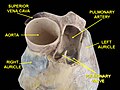
Pulmonary artery.Deep dissection.Anterior view.

CT scan of a normal lung, with different levels of pulmonary arteries.

Bronchial anatomy


See also[]
References[]
- ^ "Pulmonary Vasculature". University of Virginia School of Medicine. 2013. Retrieved 2017-06-24.
- ^ Cheitlin MD, Ursell PC (2011). "Cardiac Anatomy". In Chatterjee K (ed.). Cardiology: An Illustrated Textbook. JP Medical Ltd. p. 6. ISBN 9789350252758.
- ^ "Pulmonary Artery Anatomy". University of Virginia School of Medicine. 2013. Retrieved 2017-06-24.
- ^ Takahashi M, Fukuoka J, Nitta N, Takazakura R, Nagatani Y, Murakami Y, et al. (2008). "Imaging of pulmonary emphysema: a pictorial review". International Journal of Chronic Obstructive Pulmonary Disease. 3 (2): 193–204. doi:10.2147/COPD.S2639. PMC 2629965. PMID 18686729.
- ^ Jump up to: a b c d Schoenwolf GC, Larsen MJ, Bleyl SR, Brauer PR, Francis-West PH (2009). Larsen's human embryology (4th ed., Thoroughly rev. and updated. ed.). Philadelphia: Churchill Livingstone/Elsevier. pp. Development of the Urogenital system. ISBN 9780443068119.
- ^ Jump up to: a b Braunwald E (1992). Heart Disease: A Textbook of Cardiovascular Medicine (Fourth ed.). Philadelphia: W.B. Sanders.
- ^ "22.4 Gas Exchange – Anatomy and Physiology". opentextbc.ca. Retrieved 2019-05-22.
- ^ "Exchanging Oxygen and Carbon Dioxide - Lung and Airway Disorders". MSD Manual Consumer Version. Retrieved 2019-05-22.
- ^ Jump up to: a b c d e Colledge NR, Walker BR, Ralston SH, Britton R, eds. (2010). Davidson's Principles and Practice of Medicine (21st ed.). Edinburgh: Churchill Livingstone/Elsevier. ISBN 978-0-7020-3084-0.
- ^ "Normal Hemodynamic Parameters – Adult" (PDF). Edwards Lifesciences LLC. Archived from the original (PDF) on 2010-11-10.
- ^ Pashankar FD, Carbonella J, Bazzy-Asaad A, Friedman A (April 2008). "Prevalence and risk factors of elevated pulmonary artery pressures in children with sickle cell disease". Pediatrics. 121 (4): 777–82. doi:10.1542/peds.2007-0730. PMID 18381543. S2CID 26693444.
- ^ Gaillard F, et al. "Pulmonary hypertension". Radiopaedia. Retrieved 2017-05-27.
- ^ Piskin S, Patnaik SS, Han D, Bordones AD, Murali S, Finol EA (March 2020). "A canonical correlation analysis of the relationship between clinical attributes and patient-specific hemodynamic indices in adult pulmonary hypertension". Medical Engineering & Physics. 77: 1–9. doi:10.1016/j.medengphy.2020.01.006. PMC 7069525. PMID 32007361.
- ^ Jones J, et al. "Saddle pulmonary embolism". Radiopaedia. Retrieved 2017-10-08.
- ^ Pillalamarri NR, Patnaik SS, Piskin S, Gueldner P, Finol EA (2021-01-07). "Ex Vivo Regional Mechanical Characterization of Porcine Pulmonary Arteries". Experimental Mechanics. doi:10.1007/s11340-020-00678-2.
External links[]
- Anatomy photo:20:01-0106 at the SUNY Downstate Medical Center – "Heart: The Pericardial sac and Great vessels"
- Anatomy photo:20:07-0105 at the SUNY Downstate Medical Center – "Heart: Openings of Great Vessels into the Pericardial Sac"
- Anatomy figure: 19:05-06 at Human Anatomy Online, SUNY Downstate Medical Center – "Mediastinal surface of the right lung"
- Anatomy figure: 19:06-02 at Human Anatomy Online, SUNY Downstate Medical Center – "Mediastinal surface of the left lung"
- Histology image: 13802loa – Histology Learning System at Boston University
| show Authority control |
|---|
- Arteries of the thorax