Visual artifact

Visual artifacts (also artefacts) are anomalies apparent during visual representation as in digital graphics and other forms of imagery, especially photography and microscopy.
In digital graphics[]
- Image quality factors, different types of visual artifacts
- Compression artifacts
- Digital artifacts, visual artifacts resulting from digital image processing
- Noise
- Screen-door effect, also known as fixed-pattern noise (FPN), a visual artifact of digital projection technology
- Ghosting (television)
- Screen burn-in
- Distortion
- Silk screen effect
- Rainbow effect
- Screen tearing
- Moiré pattern
- Color banding
In video entertainment[]
Many people who use their computers as a hobby experience artifacting due to a hardware or software malfunction. The cases can differ but the usual causes are:
- Temperature issues, such as failure of cooling fan.
- Unsuited video card (graphics card) drivers.
- Drivers that have values that the graphics card is not suited with.
- Overclocking beyond the capabilities of the particular video card.
- Software bugs in the application or operating system.
The differing cases of visual artifacting can also differ between scheduled task(s).
In photography[]

These effects can occur in both analog and digital photography.
- Chromatic aberration due to optical dispersion through a lens, leading to color fringes at high-contrast boundaries in a photograph
- Motion blur
- Near-camera reflection, visual artifacts caused by the backscatter of light by unfocused particles
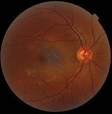
In microscopy[]

In microscopy, an artifact is an apparent structural detail that is caused by the processing of the specimen and is thus not a legitimate feature of the specimen. In light microscopy, artifacts may be produced by air bubbles trapped under the slide's cover slip.[1]
In electron microscopy, distortions may be produced in the drying out of the specimen. Staining can cause the appearance of solid chemical deposits that may be seen as structures inside the cell. Different techniques including freeze-fracturing and cell fractionation may be used to overcome the problems of artifacts.[1]
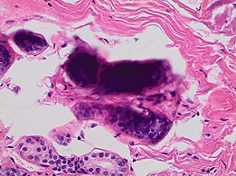
A crush artifact is an artificial elongation and distortion seen in histopathology and cytopathology studies, presumably because of iatrogenic compression of tissues. Distortion can be caused by the slightest compression of tissue and can provide difficulties in diagnosis.[2][3] It may cause chromatin to be squeezed out of nuclei.[4] Inflammatory and tumor cells are most susceptible to crush artifacts.[4]

Cellulose contamination, in H&E stain and polarized light

Cardiac muscle (bottom) with contamination from thyroid tissue (center)

Crush artifact from compression by forceps on the tissue sample
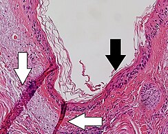
Folding artifacts (white arrows) and a crush artifact (black arrow, with cytoplasmic hypereosinophilia and nuclear pleomorphism) from a needle.

'Formalin pigment artifacts

Air bubble entrapment artifacts

Staining artifacts by residual wax, resulting in pale areas where cellular structures are not discernible.

A separation artifact in top image makes the tumor look incompletely excised, but the next microtomy level (bottom image) shows a surgical margin of connective tissue.
Stacking of cells on top of each other gives a dark look, and in this breast tissue it may mimic microcalcifications.


In radiography[]
In projectional radiography, visual artifacts that can constitute disease mimics include jewelry, clothes and skin folds.[6]

A hip fracture (black arrow) next to a skin fold (white arrow).

Bed sheets looking like lung opacities on a chest radiograph


In magnetic resonance imaging[]
In Magnetic resonance imaging, artifacts can be classified as patient-related, signal processing-dependent or hardware (machine)-related.[7]
References[]
- ^ Jump up to: a b Kent, Michael (2000). Advanced Biology (Repr. ed.). Oxford: Oxford University Press. p. 64. ISBN 0199141959.
- ^ Chatterjee, S. (September 2014). "Artefacts in histopathology". Journal of Oral and Maxillofacial Pathology. 18 (Suppl 1): S111-6. doi:10.4103/0973-029X.141346. PMC 4211218. PMID 25364159.
- ^ Komanduri S, Swanson G, Keefer L, Jakate S (December 2009). "Use of a new jumbo forceps improves tissue acquisition of Barrett's esophagus surveillance biopsies". Gastrointestinal Endoscopy. 70 (6): 1072–8.e1. doi:10.1016/j.gie.2009.04.009. PMID 19595312.
- ^ Jump up to: a b Chatterjee, Shailja (2014). "Artefacts in histopathology". Journal of Oral and Maxillofacial Pathology. 18 (4): 111. doi:10.4103/0973-029X.141346. ISSN 0973-029X. PMC 4211218.
- ^ Jump up to: a b c Taqi, SyedAhmed; Sami, SyedAbdus; Sami, LateefBegum; Zaki, SyedAhmed (2018). "A review of artifacts in histopathology". Journal of Oral and Maxillofacial Pathology. 22 (2): 279. doi:10.4103/jomfp.JOMFP_125_15. ISSN 0973-029X.
- ^ Page 46 in: Michael Darby, Nicholas Maskell, Anthony Edey, Ladli Chandratreya (2012). Pocket Tutor Chest X-Ray Interpretation. JP Medical Ltd. ISBN 9781907816062.CS1 maint: multiple names: authors list (link)
- ^ Erasmus, L.J.; Hurter, D.; Naude, M.; Kritzinger, H.G.; Acho, S. (2004). "A short overview of MRI artifacts". South African Journal of Radiology. 8 (2): 13. doi:10.4102/sajr.v8i2.127. ISSN 2078-6778. (CC-BY 4.0)
- Computer graphic artifacts
- Visual artifacts