Conjunctivochalasis
| Conjunctivochalasis | |
|---|---|
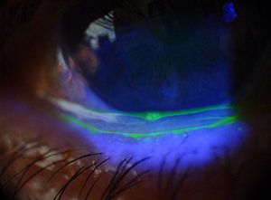 | |
| An eye with conjunctivochalasis. | |
| Specialty | Ophthalmology |
Conjunctivochalasis is a common eye surface condition characterized by the presence of excess folds of the conjunctiva located between the globe of the eye and the eyelid margin.[1]
Symptoms[]
Symptoms range from dry eye, epiphora and irritation to localized pain, foreign body sensation, subconjunctival hemorrhage and ulceration. Symptoms are often made worse by vigorous blinking.
Causes[]
Most conjunctivochalasis is thought to be caused by both a gradual thinning and stretching of the conjunctiva that accompanies age and a loss of adhesion between the conjunctiva and underlying sclera as the result of dissolution of Tenon's capsule. The resulting loose, excess conjunctiva may mechanically irritate the eye and disrupt the tear film and its outflow, leading to dry eye and excess tearing.[2] A correlation may also exist between inflammation in the eye and conjunctivochalasis, though it is unclear whether this correlation is causal.[3][4] Conjunctivochalasis may be associated with previous surgery, blepharitis, meibomian gland disorder (MGD), Ehlers-Danlos Syndrome and aqueous tear deficiency.
Diagnosis[]
Because the disorder often occurs in people with typical dry eye symptoms, it can be difficult to readily distinguish the discomfort caused by the dry eye from that directly related to the redundant conjunctiva.
Diagnosis can be made under a slit lamp upon the observation of redundant conjunctival folds. These folds can be made more apparent by staining with fluorescin dye and by applying gentle upward pressure with a finger to the eyeball through the lower lid. A tear-clearance test can also detect irregularities in the tear film.[5]
Treatment[]
Mild conjunctivochalasis can be asymptomatic and in such cases does not require treatment. Lubricating eye drops may be tried but are often ineffective.[6]
If discomfort persists after standard dry eye treatment and anti-inflammatory therapy, surgery may be undertaken to remove the conjunctival folds and restore a smooth tear film. This conjunctivoplasty surgery to correct conjunctivochalasis typically involves resection of an ellipse-shaped segment of conjunctiva just inferior to the lower lid margin, and is usually followed either by suturing or amniotic membrane graft transplantation to close the wound.[7]
References[]
- ^ Hughes WL. Conjunctivochalasis. American Journal of Ophthalmology 1942;25:48-51
- ^ Liu D. The separation of the conjunctiva can also be caused by sensitivity to chemicals suspended in the air, or to direct contact with chemicals. The reaction (in appearance) is similar to a chemical burn to the skin. Fluid builds between the conjunctiva and cornea as what appears as a blister, that looks (from a distance) like a liquid tear. Closer examination reveals the bulging conjunctiva. The stretching of the conjunctiva will remain permanent. Conjunctivochalasis A cause of Tearing and Its Management. Ophthalmic Plastic and Reconstructive Surgery 1986;2:25-28
- ^ Fodor E, Barabino S, Montaldo E, Mingari MC, Rolando M. Qualitative evaluation of ocular surface inflammation in patients with conjunctivochalasisCurr Eye Res. 2010 Aug;35(8):665-9
- ^ Erdogan-Poyraz C, Mocan MC, Bozkurt B, Gariboglu S, Irkec M, Orhan M. Elevated tear Interleukin-6 and Interleukin-8 in patients with Conjunctivochalasis Cornea. 2009 Feb;28(2):189-93.
- ^ [ Bruce A., Loughnan M.(2003) Anterior Eye and Therapeutics A-Z. Elsevier Health Sciences.]
- ^ Meller, D; Tseng, SC (1998). "Conjunctivochalasis: literature review and possible pathophysiology". Survey of Ophthalmology. 43 (3): 225–32. doi:10.1016/s0039-6257(98)00037-x. PMID 9862310.
- ^ Bosniak SL, Smith BC. Conjunctivochalasis. Advanced Ophthalmic Plastics and Reconstructive Surgery 1984;3:153-155
External links[]
- Disorders of conjunctiva