Giardia
| Giardia | |
|---|---|
| |
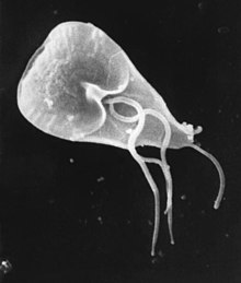
| Giardia trophozoite, SEM | |
| Scientific classification | |
| Domain: | Eukaryota
|
| (unranked): | |
| Phylum: | |
| Order: | |
| Family: | |
| Subfamily: | |
| Genus: | Giardia Künstler, 1882[1]
|
| Species | |
| |
| Synonyms | |
| |
Giardia (/dʒiːˈɑːrdiə/ or /ˈdʒɑːrdiə/) is a genus of anaerobic flagellated protozoan parasites of the phylum Metamonada that colonise and reproduce in the small intestines of several vertebrates, causing giardiasis. Their life cycle alternates between a swimming trophozoite and an infective, resistant cyst. Giardia were first described by the Dutch microscopist Antonie van Leeuwenhoek in 1681.[3] The genus is named after French zoologist Alfred Mathieu Giard.[4]
Characteristics[]
Like other diplomonads, Giardia have two nuclei, each with four associated flagella, and were thought to lack both mitochondria and a Golgi apparatus. However they are now known to possess a complex endomembrane system as well as mitochondrial remnants, called mitosomes, through mitochondrial reduction.[5] [6] [7][8] The mitosomes are not used in ATP synthesis the way mitochondria are, but are involved in the maturation of iron-sulfur proteins.[9] The synapomorphies of genus Giardia include cells with duplicate organelles, absence of cytostomes, and ventral adhesive disc.[10]
Systematics[]
About 40 species have been described from different animals, but many of them are probably synonyms.[11] Currently, five to six morphologically distinct species are recognised.[12] Giardia lamblia (=G. intestinalis, =G. duodenalis) infect humans and other mammals, G. muris is found from other mammals, G. ardeae and G. psittaci from birds, G. agilis from amphibians and G. microti from voles.[4] Other described (but not certainly valid), species include:[13]
Many different species of Giardia exist, so to differentiate between species, very specific PCR (Polymerase Chain Reactions) have been developed to detect specific Giardia spp. Gene probe-based detection is also used to differentiate between species of Giardia. A more common and less time-consuming means of identifying different species of Giardia includes microscopy and immunofluorescence techniques.[14]
Genetic and biochemical studies have revealed the heterogeneity of Giardia lamblia, which contains probably at least eight lineages or cryptic species.[15]
Genome[]
A Giardia isolate (WB) was the first diplomonad to have its genome sequenced. Its 11.7 million basepair genome is compact in structure and content with simplified basic cellular machineries and metabolism. Currently the genomes of several other Giardia isolates and diplomonads (the fish pathogens Spironucleus vortens and S. salmonicida) are being sequenced.[16]
A second isolate (the B assemblage) from humans has been sequenced along with a species from a pig (the E assemblage).[17] There are ~5000 genes in the genome. The E assemblage is more closely related to the A assemblage than is the B. A number of chromosomal rearrangements are present.
Infection[]

Giardia lives in the intestines of infected humans or other animals, individuals of which become infected by ingesting or coming into contact with contaminated foods, soil, or water tainted by the feces of an infected carrier.[18]
The symptoms of Giardia, which may begin to appear 2 days after infection, can include mild to violent diarrhoea, excess gas, stomach or abdominal cramps, upset stomach, and nausea. Resulting dehydration and nutritional loss may need immediate treatment. A typical infection can be slight, resolve without treatment, and last between 2–6 weeks, although it can sometimes last longer and/or be more severe. Coexistence with the parasite is possible (symptoms fade), but an infected individual can remain a carrier and transmit it to others. Medication containing tinidazole or metronidazole decreases symptoms and time to resolution. Albendazole is also used, and has an anthelmintic (anti-worm) property as well, ideal for certain compounded issues when a general vermicidal agent is preferred. Giardia causes a disease called giardiasis, which causes the villi of the small intestine to atrophy and flatten, resulting in malabsorption in the intestine. Lactose intolerance can persist after the eradication of Giardia from the digestive tract.[19]
Prevalence[]
Cysts are commonly detected in surface water in Russia. Water treatment plants in Moscow are occasionally contaminated with the parasites and in many cities in Russia - including Saint Petersburg - the reputation of municipal water is so bad that residents preemptively boil drinking water.[20]
See also[]
- List of parasites (human)
References[]
- ^ Künstler, J. (1882). "Sur cinq protozoaires parasites nouveaux". C. R. Acad. Sci. Paris. 95: 347–349.
- ^ Blanchard, R. (1888). "Remarques sur le megastome intestinal". Bull. Soc. Zool. Fr. 30: 18–19.
- ^ Stanley L. Erlandsen; Ernest A. Meyer (1 March 1984). Giardia and Giardiasis: Biology, Pathogenesis, and Epidemiology. Springer. pp. 131–. ISBN 978-0-306-41539-5.
- ^ a b Adam RD (July 2001). "Biology of Giardia lamblia". Clin. Microbiol. Rev. 14 (3): 447–75. doi:10.1128/CMR.14.3.447-475.2001. PMC 88984. PMID 11432808.
- ^ Tovar, Jorge; León-Avila, Gloria; Sánchez, Lidya; Sutak, Robert; Tachezy, Jan; van der Giezen, Mark; Hernández, Manuel; Müller, Miklós; Lucocq, John (2003). "Mitochondrial remnant organelles of Giardia function in iron-sulphur protein maturation". Nature. 426 (6963): 172–176. Bibcode:2003Natur.426..172T. doi:10.1038/nature01945. PMID 14614504. S2CID 4402808.
- ^ Anna Karnkowska; et al. (May 2016). "A Eukaryote without a Mitochondrial Organelle". Current Biology. 26 (10): 1274–1284. doi:10.1016/j.cub.2016.03.053. PMID 27185558.
- ^ Soltys BJ, Falah M, Gupta RS (July 1996). "Identification of endoplasmic reticulum in the primitive eukaryote Giardia lamblia using cryoelectron microscopy and antibody to Bip". J. Cell Sci. 109 (Pt 7): 1909–17. doi:10.1242/jcs.109.7.1909. PMID 8832413.
- ^ Dolezal P; Smíd O; Rada P; et al. (August 2005). "Giardia mitosomes and trichomonad hydrogenosomes share a common mode of protein targeting". Proc. Natl. Acad. Sci. U.S.A. 102 (31): 10924–9. Bibcode:2005PNAS..10210924D. doi:10.1073/pnas.0500349102. PMC 1182405. PMID 16040811.
- ^ Tovar J, et al. (2003). "Mitochondrial remnant organelles of Giardia function in iron-sulphur protein maturation". Nature. 426 (6963): 172–6. Bibcode:2003Natur.426..172T. doi:10.1038/nature01945. PMID 14614504. S2CID 4402808.
- ^ Cepicka, Ivan (September 2008). "Fornicata". Tree of Life Web Project.
- ^ Meyer E.A.; Radulescu S. (1979). "Giardia and Giardiasis". Advances in Parasitology. 17: 1–47. doi:10.1016/S0065-308X(08)60548-5. ISBN 9780120317172. PMID 395833.
no
- ^ Brusca, R.C.; Brusca, G.J. (2003). Invertebrates (2nd ed.). Sinauer Associates. ISBN 0878930973.
- ^ "Giardia Kunstler". Tree of Life Web Project. September 2008.
- ^ Mahbubani 1992
- ^ Thompson RC, Monis PT (2004). "Variation in Giardia: implications for taxonomy and epidemiology". Adv. Parasitol. Advances in Parasitology. 58: 69–137. doi:10.1016/S0065-308X(04)58002-8. ISBN 9780120317585. PMID 15603762.
- ^ Andersson, JO; et al. (2010). "The Genome of Giardia and Other Diplomonads". Anaerobic Parasitic Protozoa: Genomics and Molecular Biology. Caister Academic Press. ISBN 978-1-904455-61-5.
- ^ Jerlström-Hultqvist J, Ankarklev J, Svärd SG (2010). "Is human giardiasis caused by two different Giardia species?". Gut Microbes. 1 (6): 379–82. doi:10.4161/gmic.1.6.13608. PMC 3056102. PMID 21468219.
- ^ Filice, F.P. (1952). "Studies on the cytology and life history of a Giardia from the laboratory rat". U. C. Publications in Zoology. Berkeley CA: University of California Press. 5sex7 (2).
- ^ LaCour 2003
- ^ Henry, Laura A.; Douhovnikoff, Vladimir (2008). "Environmental Issues in Russia". Annual Review of Environment and Resources. Annual Reviews. 33 (1): 437–460. doi:10.1146/annurev.environ.33.051007.082437. ISSN 1543-5938.
External links[]
- "Giardia". Parasites. Centers for Disease Control and Prevention. March 2011.
- Mahbubani MH, Bej AK, Perlin MH, Schaefer FW, Jakubowski W, Atlas RM (January 1992). "Differentiation of Giardia duodenalis from other Giardia spp. by using polymerase chain reaction and gene probes". J. Clin. Microbiol. 30 (1): 74–8. doi:10.1128/JCM.30.1.74-78.1992. PMC 264999. PMID 1734070.
- LaCour, Michelle (2003). "Who Is Giardia?". GIARDIA. Stanford University.
- "Giardia". NCBI Taxonomy Browser. 5740.
| Microscopic discoveries 1 |
|  | ||||
|---|---|---|---|---|---|---|
| General topics |
| |||||
| Related topics |
| |||||
| Related people |
| |||||
| Recognitions |
| |||||
| ||||||
- Metamonads
- Excavata genera
- 1681 in science