Hashimoto's thyroiditis
| Hashimoto's thyroiditis | |
|---|---|
| Other names | Chronic lymphocytic thyroiditis, autoimmune thyroiditis, struma lymphomatosa, Hashimoto's disease |
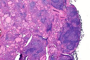 | |
| The thyroid of someone with Hashimoto's thyroiditis as seen with a microscope at low magnification | |
| Specialty | Endocrinology |
| Symptoms | Painless goiter, weight gain, feeling tired, constipation, depression, dry skin, hair loss[1] |
| Complications | Thyroid lymphoma.[2] |
| Usual onset | 30–50 years old[1][3] |
| Causes | Genetic and environmental factors.[4] |
| Risk factors | Family history, another autoimmune disease[1] |
| Diagnostic method | TSH, T4, anti-thyroid autoantibodies[1] |
| Differential diagnosis | Graves’ disease, nontoxic nodular goiter[5] |
| Treatment | Levothyroxine, surgery[1][5] |
| Frequency | 5% at some point[4] |
Hashimoto's thyroiditis, also known as chronic lymphocytic thyroiditis and Hashimoto's disease, is an autoimmune disease in which the thyroid gland is gradually destroyed.[1][6] Early on, symptoms may not be noticed.[1] Over time, the thyroid may enlarge, forming a painless goiter.[1] Some people eventually develop hypothyroidism with accompanying weight gain, fatigue, constipation, depression, hair loss, and general pains.[1] After many years the thyroid typically shrinks in size.[1] Potential complications include thyroid lymphoma.[2]
Hashimoto's thyroiditis is thought to be due to a combination of genetic and environmental factors.[4] Risk factors include a family history of the condition and having another autoimmune disease.[1] Diagnosis is confirmed with blood tests for TSH, T4, and antithyroid autoantibodies.[1] Other conditions that can produce similar symptoms include Graves’ disease and nontoxic nodular goiter.[5]
Hashimoto's thyroiditis is typically treated with levothyroxine.[1][7] If hypothyroidism is not present, some may recommend no treatment, while others may treat to try to reduce the size of the goiter.[1][8] Those affected should avoid eating large amounts of iodine; however, sufficient iodine is required especially during pregnancy.[1] Surgery is rarely required to treat the goiter.[5]
Hashimoto's thyroiditis affects about 5% of Caucasians at some point in their lives.[4] It typically begins between the ages of 30 and 50 and is much more common in women than men.[1][3] Rates of the disease appear to be increasing.[5] It was first described by the Japanese physician Hakaru Hashimoto in 1912.[9] In 1957 it was recognized as an autoimmune disorder.[10]
Signs and symptoms[]
Many symptoms are attributed to the development of Hashimoto's thyroiditis. The most common symptoms include: fatigue, weight gain, pale or puffy face, feeling cold, joint and muscle pain, constipation, dry and thinning hair, heavy menstrual flow or irregular periods, depression, panic disorder, a slowed heart rate, and problems getting pregnant and maintaining pregnancy.[11]
Hashimoto’s disease is about seven times more common in women than in men. It can occur in teens and young women, but more commonly appears in middle age, particularly for men. People who develop Hashimoto’s disease often have family members who have thyroid or other autoimmune diseases, and sometimes have other autoimmune diseases themselves.[12]
The thyroid gland may become firm, large, and lobulated in Hashimoto's thyroiditis, but changes in the thyroid can also be nonpalpable.[13] Enlargement of the thyroid is due to lymphocytic infiltration and fibrosis, rather than tissue hypertrophy. While their role in the initial destruction of the follicles is unclear, antibodies against thyroid peroxidase or thyroglobulin are relevant, as they serve as markers for detecting the disease and its severity.[14] They are thought to be the secondary products of the T cell-mediated destruction of the gland.[15]
It is also characterized by invasion of the thyroid tissue by leukocytes, mainly T-lymphocytes. A rare but serious complication is thyroid lymphoma, generally the B-cell type, non-Hodgkin lymphoma.[16]
Risk factors[]
The strong genetic component is borne out in studies on monozygotic twins, with a concordance of 38–55%, with an even higher concordance of circulating thyroid antibodies not in relation to clinical presentation (up to 80% in monozygotic twins). Neither result was seen to a similar degree in dizygotic twins, offering strong favour for high genetic aetiology.[17]
Medications that influence thyroid function[]
Certain medications or drugs have been associated with altering and interfering with thyroid function. Of these drugs, there are two main mechanisms of interference that they can have.
One of the mechanisms of interference is when a drug alters thyroid hormone serum transfer proteins.[18] Estrogen, tamoxifen, heroin, methadone, clofibrate, 5-flurouracile, mitotane, and perphenazine all increase thyroid binding globulin (TBG) concentration.[18] Androgens, anabolic steroids such as danazol, glucocorticoids, and slow release nicotinic acid all decrease TBG concentrations. Furosemide, fenoflenac, mefanamic acid, salicylates, phenytoin, diazepam, sulphonylureas, free fatty acids, and heparin all interfere with thyroid hormone binding to TBG and/or transthyretin.
The other mechanism that medications can utilize to interfere with thyroid function would be to alter extra-thryoidal metabolism of thyroid hormone. Propylthiouracil, glucocorticoids, propanolol, iondinated contrast agents, amiodarone, and clomipramine all inhibit conversion of T4 and T3.[18] Phenobarbital, rifampin, phenytoin and carbamazepine all increase hepatic metabolism.[18] Finally, cholestryamine, colestipol, aluminium hydroxide, ferrous sulphate, and sucralfate are all drugs that decrease T4 absorption or enhance excretion.[18]
HLA genes[]
The first gene locus associated with autoimmune thyroid disease was major histocompatibility complex (MHC) region on chromosome 6p21. It encodes HLAs. Specific HLA alleles have a higher affinity to autoantigenic thyroidal peptides and can contribute to autoimmune thyroid disease development. Specifically, in Hashimoto’s disease, aberrant expression of HLA II on thyrocytes has been demonstrated. They can present thyroid autoantigens and initiate autoimmune thyroid disease.[19] Susceptibility alleles are not consistent in Hashimoto’s disease. In Caucasians, various alleles are reported to be associated with the disease, including DR3, DR5 and DQ7.[20][21]
CTLA-4 genes[]
This gene is the second major immune-regulatory gene related to autoimmune thyroid disease. CTLA-4 gene polymorphisms may contribute to the reduced inhibition of T-cell proliferation and increase susceptibility to autoimmune response.[22] CTLA-4 is a major thyroid autoantibody susceptibility gene. A linkage of the CTLA-4 region to the presence of thyroid autoantibodies was demonstrated by a whole-genome linkage analysis.[23] CTLA-4 was confirmed as the main locus for thyroid autoantibodies.[24]
Protein tyrosine phosphatase nonreceptor-type 22 gene[]
PTPN22 is the most recently identified immune-regulatory gene associated with autoimmune thyroid disease. It is located on chromosome 1p13 and expressed in lymphocytes. It acts as a negative regulator of T-cell activation. Mutation in this gene is a risk factor for many autoimmune diseases. Weaker T-cell signaling may lead to impaired thymic deletion of autoreactive T cells, and increased PTPN22 function may result in inhibition of regulatory T cells, which protect against autoimmunity.[25]
[]
IFN-γ promotes cell-mediated cytotoxicity against thyroid. Mutations causing increased production of IFN-γ were associated with the severity of hypothyroidism.[26] Severe hypothyroidism is associated with mutations leading to lower production of IL-4 (Th2 cytokine suppressing cell-mediated autoimmunity),[27] lower secretion of TGF-β (inhibitor of cytokine production),[28] and mutations of FoxP3, an essential regulatory factor for the Tregs development.[29] Development of Hashimoto’s disease was associated with mutation of the gene for TNF-α (stimulator of the IFN-γ production), causing its higher concentration.[30]
Preventable environmental factors, including high iodine intake, selenium deficiency, and infectious diseases and certain drugs, have been implicated in the development of autoimmune thyroid disease in genetically predisposed individuals.[31]
Iodine[]
Excessive iodine intake is a well-established environmental factor for triggering thyroid autoimmunity. A higher prevalence of thyroid autoantibodies is in the areas with higher iodine supply. Several mechanisms by which iodine may promote thyroid autoimmunity have been proposed. Iodine exposure leads to higher iodination of thyroglobulin, increasing its immunogenicity by creating new iodine-containing epitopes or exposing cryptic epitopes. It may facilitate presentation by APC, enhance the binding affinity of the T-cell receptor, and activating specific T-cells.[32]
Iodine exposure has been shown to increase the level of reactive oxygen species. They enhance the expression of the intracellular adhesion molecule-1 on the thyroid follicular cells, which could attract the immunocompetent cells into the thyroid gland.[33]
Iodine is toxic to thyrocytes since highly reactive oxygen species may bind to membrane lipids and proteins. It causes thyrocyte damage and the release of autoantigens. Iodine also promotes follicular cell apoptosis and has an influence on immune cells (augmented maturation of dendritic cells, increased number of T cells, stimulated B-cell immunoglobulin production).[34][35]
Data from The Danish Investigation of Iodine Intake and Thyroid Disease shows that within two cohorts (males, females) with moderate and mild iodine deficiency, the levels of both thyroid peroxidase and thyroglobulin antibodies are higher in females, and prevalence rates of both antibodies increase with age.[36]
Gender[]
Study of healthy Danish twins divided to three groups (monozygotic and dizygotic same sex, and opposite sex twin pairs) estimated that genetic contribution to thyroid peroxidase antibodies susceptibility was 61% in males and 72% in females, and contribution to thyroglobulin antibodies susceptibility was 39% in males and 75% in females.[37]
The high female predominance in thyroid autoimmunity may be associated with the X chromosome. It contains sex and immune-related genes responsible for immune tolerance.[38] A higher incidence of thyroid autoimmunity was reported in patients with a higher rate of X chromosome monosomy in peripheral white blood cells.[39]
Another potential mechanism might be skewed X-chromosome inactivation, leading to the escape of X-linked self-antigens from presentation in the thymus and loss of T-cell tolerance.[citation needed]
Having other autoimmune diseases is a risk factor for developing Hashimoto’s thyroiditis, and the opposite is also true.[1] Autoimmune diseases most commonly associated to Hashimoto’s thyroiditis include celiac disease, type 1 diabetes, vitiligo, and alopecia.[40]
The genes implicated vary in different ethnic groups and the incidence is increased in people with chromosomal disorders, including Turner, Down, and Klinefelter syndromes usually associated with autoantibodies against thyroglobulin and thyroperoxidase. Progressive depletion of these cells as the cytotoxic immune response leads to higher degrees of primary hypothyroidism, presenting with low T3/T4 levels, and compensatory elevations of TSH.[citation needed]
Pathophysiology[]
Multiple mechanisms by which the pathology of Hashimoto's thyroiditis develops have been suggested.
Various autoantibodies may be present against thyroid peroxidase, thyroglobulin and TSH receptors, although a small percentage of people may have none of these antibodies present. As indicated in various twin studies, a percentage of the population may also have these antibodies without developing Hashimoto's thyroiditis. Nevertheless, antibody-dependent, cell-mediated cytotoxicity is a substantial factor behind the apoptotic fall-out of Hashimoto's thyroiditis. Activation of cytotoxic T-lymphocytes (CD8+ T-cells) in response to cell-mediated immune response affected by helper T-lymphocytes (CD4+ T-cells) is central to thyrocyte destruction. As is characteristic of type IV hypersensitivities, recruitment of macrophages is another effect of the helper T-lymphocyte activation, with Th1 axis lymphocytes producing inflammatory cytokines within thyroid tissue to further macrophage activation and migration into the thyroid gland for direct effect.[citation needed]
Gross morphological changes within the thyroid are seen in the general enlargement, which is far more locally nodular and irregular than more diffuse patterns (such as that of hyperthyroidism). While the capsule is intact and the gland itself is still distinct from surrounding tissue, microscopic examination can provide a more revealing indication of the level of damage.[41]
Histologically, the hypersensitivity is seen as diffuse parenchymal infiltration by lymphocytes, particularly plasma B-cells, which can often be seen as secondary lymphoid follicles (germinal centers, not to be confused with the normally present colloid-filled follicles that constitute the thyroid). Atrophy of the colloid bodies is lined by Hürthle cells, cells with intensely eosinophilic, granular cytoplasm, a metaplasia from the normal cuboidal cells that constitute the lining of the thyroid follicles. Severe thyroid atrophy presents often with denser fibrotic bands of collagen that remains within the confines of the thyroid capsule.[41]
Diagnosis[]
Diagnosis is usually made by detecting elevated levels of antithyroid peroxidase antibodies in the serum, but seronegative (without circulating autoantibodies) thyroiditis is also possible.[42]
Given the relatively nonspecific symptoms of initial hypothyroidism, Hashimoto's thyroiditis is often misdiagnosed as depression, cyclothymia, premenstrual syndrome, chronic fatigue syndrome, fibromyalgia, and less frequently, as erectile dysfunction or an anxiety disorder. On gross examination, a hard goiter that is not painful to the touch often presents;[41] other symptoms seen with hypothyroidism, such as periorbital myxedema, depend on the current state of progression of the response, especially given the usually gradual development of clinically relevant hypothyroidism. Testing for thyroid-stimulating hormone (TSH), free T3, free T4, and the antithyroglobulin antibodies (anti-Tg), antithyroid peroxidase antibodies (anti-TPO, or TPOAb) and antimicrosomal antibodies can help obtain an accurate diagnosis.[43] Earlier assessment of the person may present with elevated levels of thyroglobulin owing to transient thyrotoxicosis, as inflammation within the thyroid causes damage to the integrity of thyroid follicle storage of thyroglobulin; TSH secretion from the anterior pituitary increases in response to a decrease in negative feedback inhibition secondary to decreased serum thyroid hormones. Typically, T4 is the preferred thyroid hormone test for hypothyroidism.[44] This exposure of the body to substantial amounts of previously isolated thyroid enzymes is thought to contribute to the exacerbation of tolerance breakdown, giving rise to the more pronounced symptoms seen later in the disease. Lymphocytic infiltration of the thyrocyte-associated tissues often leads to the histologically significant finding of germinal center development within the thyroid gland.[citation needed]
Hashimoto's when presenting as mania is known as Prasad's syndrome after Ashok Prasad, the psychiatrist who first described it.[45]
Treatment[]
Managing hormone levels[]
Hypothyroidism caused by Hashimoto's thyroiditis is treated with thyroid hormone replacement agents such as levothyroxine, triiodothyronine, or desiccated thyroid extract. A tablet taken once a day generally keeps the thyroid hormone levels normal. In most cases, the treatment needs to be taken for the rest of the person's life. If hypothyroidism is caused by Hashimoto's thyroiditis, the TSH levels may be recommended to be kept under 3.0 mIU/l.[46]
Prognosis[]
Overt, symptomatic thyroid dysfunction is the most common complication, with about 5% of people with subclinical hypothyroidism and chronic autoimmune thyroiditis progressing to thyroid failure every year. Transient periods of thyrotoxicosis (over-activity of the thyroid) sometimes occur, and rarely the illness may progress to full hyperthyroid Graves' disease with active orbitopathy (bulging, inflamed eyes). Rare cases of fibrous autoimmune thyroiditis present with severe shortness of breath and difficulty swallowing, resembling aggressive thyroid tumors, but such symptoms always improve with surgery or corticosteroid therapy. Primary thyroid B-cell lymphoma affects fewer than one in 1000 persons, and it is more likely to affect those with long-standing autoimmune thyroiditis.[47]
Epidemiology[]
This disorder is thought to be the most common cause of primary hypothyroidism in North America; as a cause of nonendemic goiter, it is among the most common.[41] Hashimoto's thyroiditis affects about 5% of Caucasians at some point in their lives.[4] In the U.S., it affects fewer African-Americans, but is linked to greater mortality in the African-American population.[48] It is also less frequent in Asian populations.[49] About 1.0 to 1.5 in 1000 people have this disease at any time.[41] It occurs between eight and 15 times more often in women than in men. Though it may occur at any age, including in children, it is most often observed in women between 30 and 60 years of age.[47] Some research suggests a connection to the role of the placenta as an explanation for the sex difference.[50]
It is more common in regions of high iodine dietary intake, and among people who are genetically susceptible.[47]
History[]
Also known as Hashimoto's disease, Hashimoto's thyroiditis is named after Japanese physician Hakaru Hashimoto (1881−1934) of the medical school at Kyushu University,[51] who first described the symptoms of persons with struma lymphomatosa, an intense infiltration of lymphocytes within the thyroid, in 1912 in the German journal called Archiv für Klinische Chirurgie.[52][53] This paper was made up of 30 pages and 5 illustrations all describing the histological changes in the thyroid tissue. Furthermore, all results in his first study were collected from four women. These results explained the pathological characteristics observed in these women especially the infiltration of lymphoid and plasma cells as well as the formation of lymphoid follicles with germinal centers, fibrosis, degenerated thyroid epithelial cells and leukocytes in the lumen.[52] He described these traits to be histologically similar to those of Mikulic's disease. Like mentioned above, once he discovered these traits in this new disease, he names the disease struma lymphomatosa. This disease emphasized the lymphoid cell infiltration and formation of the lymphoid follicles with germinal centers, neither of which had ever been previously reported.[52]
Despite Dr.Hashimoto's discovery and publication, the disease was unfortunately not recognized as distinct from Reidel's Thyroiditis which was a common disease at that time in Europe. Although many other articles were reported and published by other researchers, Hashimoto's struma lymphomatosa was only recognized as an early phase of Reidel's Thyroiditis in the early 1900s. It was not until 1931 that the disease was recognized as a disease in its own right when researchers Allen Graham et al from Cleveland reported its symptoms and presentation in the same detailed manner as Hakuru.[52]
In 1956, Drs.Rose and Witebsky were able to demonstrate how immunization of certain rodents with extracts of other rodent's thyroid resembled the disease Hakuru and other researchers were trying to describe.[52] These doctors were also able to describe anti-thyroglobulin antibodies in blood serum samples from these same animals.
Later on in the same year, researches from the Middlesex Hospital in London were able to perform human experiments on patients who presented with similar symptoms. They purified anti-thyroglobulin antibody from their serum and were able to conclude that these sick patients have an immunological reaction to human thyroglobulin.[3] From this data, it was proposed that Hashimoto's struma could be an autoimmune disease of the thyroid gland.
In 1957, it was recognized as an autoimmune disorder and was the first organ-specific autoimmune disorder identified.[10]
Following this recognition, the same researchers from Middlesex Hospital published an article in 1962 in Lancet which ended up including a portrait of Hakuru Hashimoto.[52] The disease was able to become so much more well known from that moment and Hashimoto's disease became to appear more frequently in textbooks.
Since those discoveries, a number of autoimmune diseases have been able to be discovered with several of them having to do with thyroid-specific antibodies.
Pregnancy[]
Pregnant women who are positive for Hashimoto's thyroiditis may have decreased thyroid function or the gland may fail entirely.[54] If a woman is TPOAb-positive, clinicians can inform her of the risks for herself and her infant if the disease goes untreated. "Thyroid peroxidase antibodies (TPOAb) are detected in 10% of pregnant women," which presents risks to those pregnancies.[54] Women who have low thyroid function that has not been stabilized are at greater risk of having an infant with: low birth weight, neonatal respiratory distress, hydrocephalus, hypospadias, miscarriage, and preterm delivery.[54][55] The embryo transplantion rate and successful pregnancy outcomes are improved when Hashimoto's is treated.[55] Recommendations are to treat pregnant women only if they are TPOAb-positive throughout the entirety of their pregnancies and to screen all pregnant women for thyroid levels.[54] Close cooperation between the endocrinologist and obstetrician benefits the woman and the infant.[54][56][57] The Endocrine Society recommends screening in pregnant women who are considered high-risk for thyroid autoimmune disease.[58]
Thyroid peroxides antibodies testing is recommended for women who have ever been pregnant regardless of pregnancy outcome. "...[P]revious pregnancy plays a major role in development of autoimmune overt hypothyroidism in premenopausal women, and the number of previous pregnancies should be taken into account when evaluating the risk of hypothyroidism in a young women [sic]."[59]
Hormonal changes and trophoblast expression of key immunomodulatory molecules lead to immunosuppression and fetal tolerance. Main players in regulation of the immune response are Tregs. Both cell-mediated and humoral immune responses are attenuated, resulting in immune tolerance and suppression of autoimmunity. It has been reported that during pregnancy, levels of thyroid peroxidase and thyroglobulin antibodies decrease. After giving birth, Tregs rapidly decrease and immune responses are re-establish. It may lead to the occurrence or aggravation of the autoimmune thyroid disease.[60] In up to 50% of females with thyroid peroxidase antibodies in the early pregnancy, thyroid autoimmunity in the postpartum period exacerbates in the form of postpartum thyroiditis.[61] Higher secretion of IFN-γ and IL-4, and lower plasma cortisol concentration during pregnancy has been reported in females with postpartum thyroiditis than in healthy females. It indicates that weaker immunosuppression during pregnancy could contribute to the postpartum thyroid dysfunction.[62]
Fetal microchimerism[]
Several years after the delivery, the chimeric male cells can be detected in the maternal peripheral blood, thyroid, lung, skin, or lymph nodes. The fetal immune cells in the maternal thyroid gland may become activated and act as a trigger that may initiate or exaggerate the autoimmune thyroid disease. In Hashimoto’s disease patients, fetal microchimeric cells were detected in thyroid in significantly higher numbers than in healthy females.[63]
See also[]
- Hashimoto's encephalopathy
- Myxedematous psychosis
References[]
- ^ Jump up to: a b c d e f g h i j k l m n o p q "Hashimoto's Disease". NIDDK. May 2014. Archived from the original on 22 August 2016. Retrieved 9 August 2016.
- ^ Jump up to: a b Noureldine, SI; Tufano, RP (January 2015). "Association of Hashimoto's thyroiditis and thyroid cancer". Current Opinion in Oncology. 27 (1): 21–5. doi:10.1097/cco.0000000000000150. PMID 25390557. S2CID 32109200.
- ^ Jump up to: a b c Hiromatsu, Yuji; Satoh, Hiroshi; Amino, Nobuyuki (January 2013). "Hashimoto's Thyroiditis: History and Future Outlook". Hormones. 12 (1): 12–18. doi:10.1007/BF03401282. PMID 23624127. S2CID 38996783.
- ^ Jump up to: a b c d e Pyzik, A; Grywalska, E; Matyjaszek-Matuszek, B; Roliński, J (2015). "Immune disorders in Hashimoto's thyroiditis: what do we know so far?". Journal of Immunology Research. 2015: 979167. doi:10.1155/2015/979167. PMC 4426893. PMID 26000316.
- ^ Jump up to: a b c d e Akamizu, T; Amino, N; Feingold, KR; Anawalt, B; Boyce, A; Chrousos, G; de Herder, WW; Dungan, K; Grossman, A; Hershman, JM; Hofland, J; Kaltsas, G; Koch, C; Kopp, P; Korbonits, M; McLachlan, R; Morley, JE; New, M; Purnell, J; Singer, F; Stratakis, CA; Trence, DL; Wilson, DP (2000). "Hashimoto's Thyroiditis". In Akamizu, Takashi; Amino, Nobuyuki (eds.). Endotext. MDText. PMID 25905412.
- ^ "Hashimoto's disease". Office on Women’s Health, U.S. Department of Health and Human Services. 12 June 2017. Archived from the original on 28 July 2017. Retrieved 17 July 2017.
 This article incorporates text from this source, which is in the public domain.
- ^ "Hashimoto Thyroiditis – Endocrine and Metabolic Disorders". Merck Manuals Professional Edition. July 2016. Retrieved 30 December 2017.
- ^ "Hashimoto Thyroiditis – Hormonal and Metabolic Disorders". Merck Manuals Consumer Version. Retrieved 30 December 2017.
- ^ Shoenfeld, Yehuda; Cervera, Ricard; Gershwin, M. Eric (2010). Diagnostic Criteria in Autoimmune Diseases. Springer Science & Business Media. p. 216. ISBN 978-1-60327-285-8.
- ^ Jump up to: a b Moore, Elaine A.; Wilkinson, Samantha (2009). The Promise of Low Dose Naltrexone Therapy: Potential Benefits in Cancer, Autoimmune, Neurological and Infectious Disorders. McFarland. p. 30. ISBN 978-0-7864-5258-3.
- ^ "Hashimoto's disease – Symptoms and causes". Mayo Clinic. Retrieved 5 October 2018.
- ^ "Hashimoto's disease fact sheet". Office on Women's Health, U.S. Department of Health and Human Services, womenshealth.gov (or girlshealth.gov). 16 July 2012. Archived from the original on 2 December 2014. Retrieved 23 November 2014.
- ^ Page 56 in: Staecker, Hinrich; Thomas R. Van De Water; Van de Water, Thomas R. (2006). Otolaryngology: basic science and clinical review. Stuttgart: Thieme. ISBN 978-0-86577-901-3.
- ^ "Pathogenesis of Hashimoto's thyroiditis (chronic autoimmune thyroiditis)". UpToDate.
- ^ "Hashimoto Thyroiditis". NCBI StatPearls. 2019.
- ^ Dayan, Colin M.; Daniels, Gilbert H. (11 July 1996). "Chronic Autoimmune Thyroiditis". New England Journal of Medicine. 335 (2): 99–107. doi:10.1056/NEJM199607113350206. PMID 8649497.
- ^ Chistiakov, Dimitry A (2005). "Immunogenetics of Hashimoto's thyroiditis". Journal of Autoimmune Diseases. 2 (1): 1. doi:10.1186/1740-2557-2-1. PMC 555850. PMID 15762980.
- ^ Jump up to: a b c d e Surks, Martin I.; Sievert, Rubens (21 December 1995). Wood, Alastair J.J. (ed.). "Drugs and Thyroid Function". New England Journal of Medicine. 333 (25): 1688–1694. doi:10.1056/NEJM199512213332507. ISSN 0028-4793. PMID 7477223.
- ^ Jacobson, Eric M.; Huber, Amanda; Tomer, Yaron (2008). "The HLA gene complex in thyroid autoimmunity: From epidemiology to etiology". Journal of Autoimmunity. 30 (1–2): 58–62. doi:10.1016/j.jaut.2007.11.010. PMC 2244911. PMID 18178059.
- ^ Tendon, N.; Zhang, L.; Weetman, A. P. (May 1991). "HLA Associations with Hashimoto's thyroiditis". Clinical Endocrinology. 34 (5): 383–386. doi:10.1111/j.1365-2265.1991.tb00309.x. PMID 1676351. S2CID 28987581.
- ^ Bogner, U.; Badenhoop, K.; Peters, H.; Schmieg, D.; Mayr, W. R.; Usadel, K. H.; Schleusener, H. (January 1992). "Hla-Dr/Dq Gene Variation in Nongoitrous Autoimmune Thyroiditis at the Serological and Molecular Level". Autoimmunity. 14 (2): 155–158. doi:10.3109/08916939209083135. PMID 1363895.
- ^ Zaletel, Katja; Gaberšček, Simona (December 2011). "Hashimoto's Thyroiditis: From Genes to the Disease". Current Genomics. 12 (8): 576–588. doi:10.2174/138920211798120763. PMC 3271310. PMID 22654557.
- ^ Tomer, Yaron; Greenberg, David A.; Barbesino, Giuseppe; Concepcion, Erlinda; Davies, Terry F. (March 2001). "CTLA-4 and Not CD28 Is a Susceptibility Gene for Thyroid Autoantibody Production 1". The Journal of Clinical Endocrinology & Metabolism. 86 (4): 1687–1693. doi:10.1210/jcem.86.4.7372. PMID 11297604.
- ^ Ban, Y; Davies, T F; Greenberg, D A; Kissin, A; Marder, B; Murphy, B; Concepcion, E S; Villanueva, R B; Barbesino, G; Ling, V; Tomer, Y (December 2003). "Analysis of the CTLA-4, CD28, and inducible costimulator (ICOS) genes in autoimmune thyroid disease". Genes & Immunity. 4 (8): 586–593. doi:10.1038/sj.gene.6364018. PMID 14647199. S2CID 6920190.
- ^ Burn, Garth L.; Svensson, Lena; Sanchez-Blanco, Cristina; Saini, Manoj; Cope, Andrew P. (1 December 2011). "Why is PTPN22 a good candidate susceptibility gene for autoimmune disease?". FEBS Letters. 585 (23): 3689–3698. doi:10.1016/j.febslet.2011.04.032. PMID 21515266. S2CID 21572847.
- ^ Ito, Chisato; Watanabe, Mikio; Okuda, Noriko; Watanabe, Chikami; Iwatani, Yoshinori (2006). "Association between the Severity of Hashimoto's Disease and the Functional +874A/T Polymorphism in the Interferon-.GAMMA. Gene". Endocrine Journal. 53 (4): 473–478. doi:10.1507/endocrj.k06-015. PMID 16820703.
- ^ Nanba, Takashi; Watanabe, Mikio; Akamizu, Takashi; Iwatani, Yoshinori (1 March 2008). "The −590CC Genotype in the IL4 Gene as a Strong Predictive Factor for the Development of Hypothyroidism in Hashimoto Disease". Clinical Chemistry. 54 (3): 621–623. doi:10.1373/clinchem.2007.099739. PMID 18310157.
- ^ Yamada, H.; Watanabe, M.; Nanba, T.; Akamizu, T.; Iwatani, Y. (March 2008). "The +869T/C polymorphism in the transforming growth factor-β1 gene is associated with the severity and intractability of autoimmune thyroid disease: TGF-β1 SNP and autoimmune thyroid disease". Clinical & Experimental Immunology. 151 (3): 379–382. doi:10.1111/j.1365-2249.2007.03575.x. PMC 2276968. PMID 18190611.
- ^ Inoue, N.; Watanabe, M.; Morita, M.; Tomizawa, R.; Akamizu, T.; Tatsumi, K.; Hidaka, Y.; Iwatani, Y. (December 2010). "Association of functional polymorphisms related to the transcriptional level of FOXP3 with prognosis of autoimmune thyroid diseases: FoxP3 SNP and autoimmune thyroid disease". Clinical & Experimental Immunology. 162 (3): 402–406. doi:10.1111/j.1365-2249.2010.04229.x. PMC 3026543. PMID 20942809.
- ^ Inoue, N.; Watanabe, M.; Nanba, T.; Wada, M.; Akamizu, T.; Iwatani, Y. (May 2009). "Involvement of functional polymorphisms in the TNFA gene in the pathogenesis of autoimmune thyroid diseases and production of anti-thyrotropin receptor antibody". Clinical & Experimental Immunology. 156 (2): 199–204. doi:10.1111/j.1365-2249.2009.03884.x. PMC 2759465. PMID 19250279.
- ^ Saranac, L.; Zivanovic, S.; Bjelakovic, B.; Stamenkovic, H.; Novak, M.; Kamenov, B. (2011). "Why Is the Thyroid So Prone to Autoimmune Disease". Hormone Research in Paediatrics. 75 (3): 157–165. doi:10.1159/000324442. PMID 21346360.
- ^ Rose, Noel R.; Bonita, Raphael; Burek, C.Lynne (February 2002). "Iodine: an environmental trigger of thyroiditis". Autoimmunity Reviews. 1 (1–2): 97–103. CiteSeerX 10.1.1.326.5700. doi:10.1016/s1568-9972(01)00016-7. PMID 12849065.
- ^ Burek, C. Lynne; Talor, Monica V. (November 2009). "Environmental triggers of autoimmune thyroiditis". Journal of Autoimmunity. 33 (3–4): 183–189. doi:10.1016/j.jaut.2009.09.001. PMC 2790188. PMID 19818584.
- ^ Fountoulakis, S; Philippou, G; Tsatsoulis, A (January 2007). "The role of iodine in the evolution of thyroid disease in Greece: from endemic goiter to thyroid autoimmunity". Hormones. 6 (1): 25–35. PMID 17324915.
- ^ Yu, Xiujie; Li, Lanying; Li, Qingxin; Zang, Xiaoyi; Liu, Zebing (November 2011). "TRAIL and DR5 Promote Thyroid Follicular Cell Apoptosis in Iodine Excess-Induced Experimental Autoimmune Thyroiditis in NOD Mice". Biological Trace Element Research. 143 (2): 1064–1076. doi:10.1007/s12011-010-8941-5. PMID 21225479. S2CID 10926594.
- ^ Pedersen, Inge Bülow; Knudsen, Nils; Jørgensen, Torben; Perrild, Hans; Ovesen, Lars; Laurberg, Peter (January 2003). "Thyroid peroxidase and thyroglobulin autoantibodies in a large survey of populations with mild and moderate iodine deficiency: Thyroid autoantibodies and iodine deficiency". Clinical Endocrinology. 58 (1): 36–42. doi:10.1046/j.1365-2265.2003.01633.x. PMID 12519410. S2CID 23758580.
- ^ Hansen, Pia S; Brix, Thomas H; Iachine, Ivan; Kyvik, Kirsten O; Hegedüs, Laszlo (January 2006). "The relative importance of genetic and environmental effects for the early stages of thyroid autoimmunity: a study of healthy Danish twins". European Journal of Endocrinology. 154 (1): 29–38. doi:10.1530/eje.1.02060. PMID 16381988.
- ^ McCombe, P. A.; Mackay, J. M. Greer and I. R.; Mackay, IR (30 November 2009). "Sexual Dimorphism in Autoimmune Disease". Current Molecular Medicine. 9 (9): 1058–1079. doi:10.2174/156652409789839116. PMID 19747114.
- ^ Invernizzi, Pietro; Miozzo, Monica; Selmi, Carlo; Persani, Luca; Battezzati, Pier Maria; Zuin, Massimo; Lucchi, Simona; Meroni, Pier Luigi; Marasini, Bianca; Zeni, Silvana; Watnik, Mitchell; Grati, Francesca R.; Simoni, Giuseppe; Gershwin, M. Eric; Podda, Mauro (1 July 2005). "X Chromosome Monosomy: A Common Mechanism for Autoimmune Diseases". The Journal of Immunology. 175 (1): 575–578. doi:10.4049/jimmunol.175.1.575. PMID 15972694. S2CID 40557667.
- ^ Radetti, Giorgio (2014). "Clinical Aspects of Hashimoto's Thyroiditis". Paediatric Thyroidology. Endocrine Development. 26. pp. 158–170. doi:10.1159/000363162. ISBN 978-3-318-02720-4. PMID 25231451.
- ^ Jump up to: a b c d e Maitra, Anirban (2014). "The Endocrine System". In Kumar, Vinay; Abbas, Abul K.; Aster, Jon C. (eds.). Robbins and Cotran Pathologic Basis of Disease. Elsevier Health Sciences. pp. 1073–1140. ISBN 978-0-323-29635-9.
- ^ Grani, Giorgio; Carbotta, Giovanni; Nesca, Angela; D’Alessandri, Mimma; Vitale, Martina; Del Sordo, Marianna; Fumarola, Angela (June 2015). "A comprehensive score to diagnose Hashimoto's thyroiditis: a proposal". Endocrine. 49 (2): 361–365. doi:10.1007/s12020-014-0441-5. PMID 25280964. S2CID 23026213.
- ^ Giannini, AJ (1986). The Biological Foundations of Clinical Psychiatry. New Hyde Park, NY: Medical Examination Publishing Company. pp. 193–198. ISBN 978-0-87488-449-4.
- ^ Hashimoto Thyroiditis~workup at eMedicine
- ^ Weiner, M. J.; Kennedy, C. (March 1988). "Prasad's Syndrome". British Journal of Psychiatry. 152 (3): 438–439. doi:10.1192/bjp.152.3.438b. PMID 3167392.
- ^ "Does Your Doctor Know About the New TSH Lab Standards?". Archived from the original on 4 December 2010.
- ^ Jump up to: a b c Fabrizio Monaco (2012). Thyroid Diseases. Taylor and Francis. p. 78. ISBN 978-1-4398-6839-3.
- ^ Boyles, Salynn (23 May 2013). "Hypothyroidism Hikes Death Risk in Blacks". MedPage Today.
- ^ McLeod, Donald S. A.; Caturegli, Patrizio; Cooper, David S.; Matos, Peter G.; Hutfless, Susan (16 April 2014). "Variation in Rates of Autoimmune Thyroid Disease by Race/Ethnicity in US Military Personnel". JAMA. 311 (15): 1563–1565. doi:10.1001/jama.2013.285606. PMID 24737370. Lay summary.
- ^ Natri, Heini; Garcia, Angela R.; Buetow, Kenneth H.; Trumble, Benjamin C.; Wilson, Melissa A. (July 2019). "The Pregnancy Pickle: Evolved Immune Compensation Due to Pregnancy Underlies Sex Differences in Human Diseases". Trends in Genetics. 35 (7): 478–488. doi:10.1016/j.tig.2019.04.008. PMC 6611699. PMID 31200807.
- ^ Hakaru Hashimoto at Who Named It?
- ^ Jump up to: a b c d e f Hiromatsu, Yuji; Satoh, Hiroshi; Amino, Nobuyuki (January 2013). "Hashimoto's Thyroiditis: History and Future Outlook". Hormones. 12 (1): 12–18. doi:10.1007/BF03401282. ISSN 1109-3099. PMID 23624127. S2CID 38996783.
- ^ Hashimoto, Hakaru (1912). "Zur Kenntnis der lymphomatösen Veränderung der Schilddrüse (Struma lymphomatosa)" [Knowledge of lymphomatous changes in the thyroid gland (goiter lymphomatosa)]. Archiv für Klinische Chirurgie (in German). 97: 219–248. NAID 10005555208.
- ^ Jump up to: a b c d e Lepoutre, Thibault; Debiève, Frederic; Gruson, Damien; Daumerie, Chantal (1 January 2012). "Reduction of Miscarriages through Universal Screening and Treatment of Thyroid Autoimmune Diseases". Gynecologic and Obstetric Investigation. 74 (4): 265–273. doi:10.1159/000343759. PMID 23147711.
- ^ Jump up to: a b Gaberšček, Simona; Zaletel, Katja (September 2011). "Thyroid physiology and autoimmunity in pregnancy and after delivery". Expert Review of Clinical Immunology. 7 (5): 697–707. doi:10.1586/eci.11.42. PMID 21895480.
- ^ Budenhofer, Brigitte K.; Ditsch, Nina; Jeschke, Udo; Gärtner, Roland; Toth, Bettina (January 2013). "Thyroid (dys-)function in normal and disturbed pregnancy". Archives of Gynecology and Obstetrics. 287 (1): 1–7. doi:10.1007/s00404-012-2592-z. PMID 23104052. S2CID 24969196.
- ^ Balucan, Francis S.; Morshed, Syed A.; Davies, Terry F. (2013). "Thyroid Autoantibodies in Pregnancy: Their Role, Regulation and Clinical Relevance". Journal of Thyroid Research. 2013: 182472. doi:10.1155/2013/182472. PMC 3652173. PMID 23691429.
- ^ "Endocrine Experts Support Screening for Thyroid Dysfunction in Pregnant Women". Endocrine Society. Endocrine Society. 26 March 2015. Archived from the original on 8 October 2015. Retrieved 4 October 2015.
- ^ Carlé, Allan; Pedersen, Inge Bülow; Knudsen, Nils; Perrild, Hans; Ovesen, Lars; Rasmussen, Lone Banke; Laurberg, Peter (1 June 2014). "Development of Autoimmune Overt Hypothyroidism Is Highly Associated With Live Births and Induced Abortions but Only in Premenopausal Women". The Journal of Clinical Endocrinology & Metabolism. 99 (6): 2241–2249. doi:10.1210/jc.2013-4474. PMID 24694338.
- ^ Weetman, Anthony P. (June 2010). "Immunity, thyroid function and pregnancy: molecular mechanisms". Nature Reviews Endocrinology. 6 (6): 311–318. doi:10.1038/nrendo.2010.46. PMID 20421883. S2CID 9900120.
- ^ Lazarus, John H. (1 March 2011). "The Continuing Saga of Postpartum Thyroiditis". The Journal of Clinical Endocrinology & Metabolism. 96 (3): 614–616. doi:10.1210/jc.2011-0091. PMID 21378224.
- ^ Kokandi, A. A.; Parkes, A. B.; Premawardhana, L. D. K. E.; John, R.; Lazarus, J. H. (March 2003). "Association of Postpartum Thyroid Dysfunction with Antepartum Hormonal and Immunological Changes". The Journal of Clinical Endocrinology & Metabolism. 88 (3): 1126–1132. doi:10.1210/jc.2002-021219. PMID 12629095.
- ^ Koopmans, Marije; Kremer Hovinga, Idske C.L.; Baelde, Hans J.; Harvey, Mark S.; de Heer, Emile; Bruijn, Jan A.; Bajema, Ingeborg M. (June 2008). "Chimerism occurs in thyroid, lung, skin and lymph nodes of women with sons". Journal of Reproductive Immunology. 78 (1): 68–75. doi:10.1016/j.jri.2008.01.002. PMID 18329105.
| Classification | |
|---|---|
| External resources |
| show Authority control |
|---|
- Aging-associated diseases
- Autoimmune diseases
- Endocrine diseases
- Thyroid disease