Lichen nitidus
This article may be too technical for most readers to understand. (October 2012) |
| Lichen nitidus | |
|---|---|
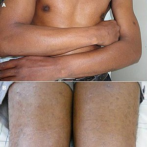 | |
| Photography of lichen nitidus: A) Glistening forearm papules; B) Papular lesions of lower limbs | |
| Specialty | Dermatology |
Lichen nitidus is a chronic inflammatory disease of unknown cause [1] characterized by 1–2 mm, discrete and uniform, shiny, flat-topped, pale flesh-colored or reddish-brown papules[2][3] that may appear as hypopigmented against dark skin. Occasionally, minimal scaling is present or can be induced by rubbing the surface of the papules.[3] The disease usually affects children and young adults[4] and is painless and usually nonpruritic, although protracted itching may occur in some cases.[3][5] It is sometimes referred to by dermatologists as "mini lichen planus".
Presentation[]
Linear arrangements of these papules is common (referred to as a Koebner phenomenon), especially on the forearms,[2][5] but may occasionally be grouped, though not confluent, on flexural areas.[2] Generally, the initial lesions are localized, and remain so, to the chest, abdomen, glans penis, and flexor aspects of the upper extremities;[6] however, less commonly, the disease process can (1) be strictly isolated to the palms and soles,[7] presenting with many hyperkeratotic, yellow papules that may coalesce into plaques that fissure[3][7] or “...sometimes a non-specific keratoderma resembling chronic eczema,”[7] or (2) become more widespread, with papules widely distributed on the body—the extensor surfaces of the elbows, wrists, and hands, folds of the neck, submammary region in females, groin, thighs, ankles, and feet[1][2]—and fusing into erythematous, minimally scaled plaques, with redness that develops tints of violet, brown, and yellow.[3][4]
Pathology[]

The histology of lichen nitidus is significant for a "...localized granulomatous lymphohistiocytic infiltrate in an expanded dermal papilla with thinning of overlying epidermis and downward extension of the rete ridges at the lateral margin of the infiltrate, producing a typical 'claw clutching a ball' picture...."[1]
Treatment[]
Generally, lichen nitidus is asymptomatic and self-limited; therefore, no treatment is required. However, if persistent pruritus is present, or the appearance “...interferes with daily activities or outlook...”[2] topical glucocorticoids may be tried. If the disease process is symptomatic, generalized and extensive, oral glucocorticoids may be indicated.[2] Other reported treatments include PUVA, UVA/UVB phototherapy,[6] astemizole,[1] acitretin, and etretinate.[2] When appears with sun/humidity; air conditioning (cool dry air) reduces swelling and discomfort.
See also[]
- Lichen planus
- List of cutaneous conditions
References[]
- ^ Jump up to: a b c d Al-Mutairi N, Hassanein A, Nour-Eldin O, Arun J (2005). "Generalized lichen nitidus". Pediatr Dermatol. 22 (2): 158–60. doi:10.1111/j.1525-1470.2005.22215.x. PMID 15804308.
- ^ Jump up to: a b c d e f g Berger, Timothy G.; Odom, Richard B.; Andrews, George E.; James, William D. (2000). Andrews' Diseases of the skin: clinical dermatology. Philadelphia: W. B. Saunders. pp. 277–80. ISBN 0-7216-5832-6.
- ^ Jump up to: a b c d e Fitzpatrick, Thomas B.; Freedberg, Irwin M. (1999). Fitzpatrick's dermatology in general medicine. New York: McGraw-Hill, Health Professions Division. pp. 577–81. ISBN 0-07-912938-2.
- ^ Jump up to: a b Soroush V, Gurevitch AW, Peng SK (1999). "Generalized lichen nitidus: case report and literature review". Cutis. 64 (2): 135–6. PMID 10467510.
- ^ Jump up to: a b Maeda M (1994). "A case of generalized lichen nitidus with Koebner's phenomenon". J. Dermatol. 21 (4): 273–7. doi:10.1111/j.1346-8138.1994.tb01736.x. PMID 8056902.
- ^ Jump up to: a b Do MO, Kim MJ, Kim SH, Myung KB, Choi YW (2007). "Generalized Lichen Nitidus Successfully Treated with Narrow-band UVB Phototherapy : Two Cases Report". J. Korean Med. Sci. 22 (1): 163–6. doi:10.3346/jkms.2007.22.1.163. PMC 2693559. PMID 17297274.[dead link]
- ^ Jump up to: a b c Thibaudeau A, Maillard H, Croué A, Belperron P, Avenel Audran M, Verret JL (2004). "[Palmoplantar lichen nitidus: a rare cause of palmoplantar hyperkeratosis.]". Ann Dermatol Venereol (in French). 131 (8–9): 822–4. doi:10.1016/S0151-9638(04)93769-6. PMID 15505553.
External links[]
| Classification | |
|---|---|
| External resources |
- Lichen Nitidus at DermNet photo library
- Lichenoid eruptions
- Papulosquamous disorders