Precancerous condition
This article relies largely or entirely on a single source. (July 2012) |
| Precancerous condition | |
|---|---|
| Other names | Premalignant condition, precancer, premalignancy, dysplasia, intraepithelial neoplasm, carcinoma in situ |
 | |
| Micrograph of high grade squamous intraepithelial lesion, a precancerous condition of the uterine cervix. Pap stain. | |
| Specialty | Oncology |
A precancerous condition is a condition or lesion involving abnormal cells which are associated with an increased risk of developing into cancer.[1][2][3] Clinically, precancerous conditions encompass a variety of conditions or lesions with an increased risk of developing into cancer. Some of the most common precancerous conditions include certain colon polyps, which can progress into colon cancer, monoclonal gammopathy of undetermined significance, which can progress into multiple myeloma or myelodysplastic syndrome.[4] and cervical dysplasia, which can progress into cervical cancer.[5] Bronchial premalignant lesions can progress to squamous cell carcinoma of the lung.[6]
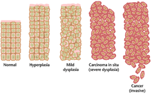
Pathologically, precancerous lesions can range from benign neoplasias, which are tumors which do not invade neighboring normal tissues or spread to distant organs, to dysplasia,[1] a collection of abnormal cells which, in some cases, has an increased risk of progressing to anaplasia and invasive cancer. Sometimes, the term "precancer" is also used for carcinoma in situ, which is a noninvasive cancer that has not progressed to an aggressive, invasive stage. As with other precancerous conditions, not all carcinoma in situ will become an invasive disease.
Classification[]
The term precancerous or premalignant condition may refer to certain conditions, such as monoclonal gammopathy of unknown significance, or to certain lesions, such as colorectal adenoma (colon polyps), which have the potential to progress into cancer (see: Malignant transformation). Premalignant lesions are morphologically atypical tissue which appear abnormal when viewed under the microscope, and which are more likely to progress to cancer than normal tissue.[7] Precancerous conditions and lesions affect a variety of organ systems, including the skin, oral cavity, stomach, colon, lung, and hematological system. Some authorities also refer to hereditary genetic conditions which predispose to developing cancer, such as hereditary nonpolyposis colorectal cancer, as a precancerous condition, as individuals with these conditions have a much higher risk of developing cancer in certain organs.[3]
Signs and symptoms[]
The signs and symptoms of precancerous conditions differ based on the organ affected. In many cases, individuals with precancerous conditions do not experience any symptoms. Precancerous conditions of the skin or oral cavity often appear as visible lesions without associated pain or discomfort,[7] while precancerous conditions of the hematological system are typically asymptomatic, or in the case of monoclonal gammopathy of unknown significance, may rarely cause numbness and tingling in the hands and feet or difficulty with balance[8] (see: peripheral neuropathy).
Causes[]
In many cases, risk factors for precancerous conditions and lesions are the same risk factors that predispose individuals to a specific cancer. For example, individuals with cervical or anal infection with oncogenic, or cancer causing, strains of human papilloma virus (HPV) are at elevated risk for cervical and anal cancers,[9] respectively, as well as for cervical and anal dysplasia.[9] Similarly, sun exposure is an important risk factor for both actinic keratosis[10] as well as skin cancer. Smoking is a risk factor for premalignant (as well as malignant) lung lesions. Hereditary conditions that predispose to cancer can also predispose to premalignant lesions. However, in many cases, precancerous conditions or lesions can be sporadic and idiopathic in nature, meaning that they are not associated with a hereditary genetic predisposition to the particular cancer, nor with a direct causative agent or other identifiable cause.[11]
Pathophysiology[]
The pathophysiology of precancerous lesions is thought to be similar to that of cancer, and varies depending on the disease site and type of lesion.[12] It is thought that cancer is preceded by a clinically silent premalignant phase during which oncogenic genetic and epigenetic alterations accumulate. The duration of this premalignant phase can vary from cancer to cancer and from individual to individual.[11] Increasing evidence suggests that evasion of the immune system occurs in premalignant lesions,[13] and that the nature of the immune response to these lesions may determine if they progress to cancer or regress.[14][15]
Examples[]
Skin[]
- actinic keratosis[16]
- Bowen's disease (intraepidermal carcinoma/squamous carcinoma in situ)
- dyskeratosis congenita
Breast[]
- ductal carcinoma in situ
- Sclerosing adenosis
- Small duct papilloma
Head and neck/oral[]
- oral submucous fibrosis
- erythroplakia
- lichen planus (oral)
- leukoplakia
- proliferative verrucous leukoplakia[7]
- stomatitis nicotina[17]
Gastrointestinal[]
- Barrett's esophagus
- atrophic gastritis
- colon polyp
- Plummer-Vinson syndrome (sideropenic dysphagia)[7]
- hereditary nonpolyposis colorectal cancer[7]
- Ulcerative colitis
- Crohn's disease
Respiratory
- Bronchial premalignant lesions
Gynecological[]
- cervical dysplasia (cervical intraepithelial neoplasm, CIN)
- vaginal intraepithelial neoplasm (VAIN)[18]
- anal dysplasia (also see: anal cancer)
- lichen sclerosus
- Bowen's disease (penile or vulvar)[19]
- erythroplasia of Queyrat[19]
Urological[]
Hematological[]
- monoclonal gammopathy of unknown significance
References[]
- ^ a b "NCI Dictionary of Cancer Terms". National Cancer Institute. 2011-02-02. Retrieved 2018-03-28.
- ^ "Precancerous conditions of the colon or rectum". Canadian cancer society. Retrieved 2018-03-19.
- ^ a b "Precancerous conditions of the esophagus". Canadian cancer society. Retrieved 2018-03-19.
- ^ Korde N, Kristinsson SY, Landgren O (May 2011). "Monoclonal gammopathy of undetermined significance (MGUS) and smoldering multiple myeloma (SMM): novel biological insights and development of early treatment strategies". Blood. 117 (21): 5573–81. doi:10.1182/blood-2011-01-270140. PMC 3316455. PMID 21441462.
- ^ "Precancerous conditions of the cervix". Canadian cancer society. Retrieved 2018-03-19.
- ^ Beane, Jennifer; Mazzilli, Sarah A.; Tassinari, Anna M.; Liu, Gang; Zhang, Xiaohui; Liu, Hanqiao; Buncio, Anne Dy; Dhillon, Samjot S.; Platero, Suso J.; Lenburg, Marc E.; Reid, Mary E. (2017-09-01). "Detecting the Presence and Progression of Premalignant Lung Lesions via Airway Gene Expression". Clinical Cancer Research. 23 (17): 5091–5100. doi:10.1158/1078-0432.CCR-16-2540. ISSN 1557-3265. PMC 7404813. PMID 28533227.
- ^ a b c d e Yardimci G, Kutlubay Z, Engin B, Tuzun Y (December 2014). "Precancerous lesions of oral mucosa". World Journal of Clinical Cases. 2 (12): 866–72. doi:10.12998/wjcc.v2.i12.866. PMC 4266835. PMID 25516862.
- ^ "MGUS - MGUS Multiple Myeloma - MGUS Myeloma -Monoclonal Gammopathy". Multiple Myeloma Research Foundation. Archived from the original on 2017-07-10. Retrieved 2018-03-28.
- ^ a b Roberts JR, Siekas LL, Kaz AM (February 2017). "Anal intraepithelial neoplasia: A review of diagnosis and management". World Journal of Gastrointestinal Oncology. 9 (2): 50–61. doi:10.4251/wjgo.v9.i2.50. PMC 5314201. PMID 28255426.
- ^ "Actinic keratosis - Symptoms and causes". Mayo Clinic. Retrieved 2018-03-28.
- ^ a b Willimsky G, Czéh M, Loddenkemper C, Gellermann J, Schmidt K, Wust P, Stein H, Blankenstein T (July 2008). "Immunogenicity of premalignant lesions is the primary cause of general cytotoxic T lymphocyte unresponsiveness". The Journal of Experimental Medicine. 205 (7): 1687–700. doi:10.1084/jem.20072016. PMC 2442645. PMID 18573907.
- ^ Hyndman IJ (April 2016). "Review: the Contribution of both Nature and Nurture to Carcinogenesis and Progression in Solid Tumours". Cancer Microenvironment. 9 (1): 63–9. doi:10.1007/s12307-016-0183-4. PMC 4842185. PMID 27066794.
- ^ Mascaux, Céline; Angelova, Mihaela; Vasaturo, Angela; Beane, Jennifer; Hijazi, Kahkeshan; Anthoine, Geraldine; Buttard, Bénédicte; Rothe, Françoise; Willard-Gallo, Karen; Haller, Annick; Ninane, Vincent (July 2019). "Immune evasion before tumour invasion in early lung squamous carcinogenesis". Nature. 571 (7766): 570–575. doi:10.1038/s41586-019-1330-0. ISSN 1476-4687. PMID 31243362. S2CID 195657244.
- ^ Maoz, Asaf; Merenstein, Carter; Koga, Yusuke; Potter, Austin; Gower, Adam C.; Liu, Gang; Zhang, Sherry; Liu, Hanqiao; Stevenson, Christopher; Spira, Avrum; Reid, Mary E. (September 2021). "Elevated T cell repertoire diversity is associated with progression of lung squamous cell premalignant lesions". Journal for Immunotherapy of Cancer. 9 (9): e002647. doi:10.1136/jitc-2021-002647. ISSN 2051-1426. PMC 8477334. PMID 34580161.
- ^ Beane, Jennifer E.; Mazzilli, Sarah A.; Campbell, Joshua D.; Duclos, Grant; Krysan, Kostyantyn; Moy, Christopher; Perdomo, Catalina; Schaffer, Michael; Liu, Gang; Zhang, Sherry; Liu, Hanqiao (2019-04-23). "Molecular subtyping reveals immune alterations associated with progression of bronchial premalignant lesions". Nature Communications. 10 (1): 1856. Bibcode:2019NatCo..10.1856B. doi:10.1038/s41467-019-09834-2. ISSN 2041-1723. PMC 6478943. PMID 31015447.
- ^ "Actinic Keratosis". skincancer.org. Retrieved 2018-03-19.
- ^ Neville BW, Day TA (July 2002). "Oral cancer and precancerous lesions". CA: A Cancer Journal for Clinicians. 52 (4): 195–215. doi:10.3322/canjclin.52.4.195. PMID 12139232. S2CID 3238352.
- ^ "What Is Vaginal Cancer?". www.cancer.org. Retrieved 2018-03-28.
- ^ a b Arya M, Kalsi J, Kelly J, Muneer A (March 2013). "Malignant and premalignant lesions of the penis". BMJ. 346: f1149. doi:10.1136/bmj.f1149. PMID 23468288. S2CID 33771829.
- ^ "Bladder Cancer Staging | Bladder Cancer Stages". www.cancer.org. Retrieved 2018-03-28.
External links[]
| Look up premalignant or precancerous in Wiktionary, the free dictionary. |
- Oncology
- Medical terminology