Pseudostratified columnar epithelium
This article needs additional citations for verification. (January 2015) |
| Pseudostratified columnar epithelium | |
|---|---|
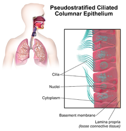 Illustration depicting Pseudostratified Ciliated Columnar Epithelium. | |
| Details | |
| Function | Epithelium |
| Identifiers | |
| TH | H2.00.02.0.02021 |
| FMA | 45572 |
| Anatomical terms of microanatomy | |
| This article is part of a series on |
| Epithelia |
|---|
| Squamous epithelial cell |
| Columnar epithelial cell |
| Cuboidal epithelial cell |
| Specialised epithelia |
|
| Other |

A pseudostratified epithelium is a type of epithelium that, though comprising only a single layer of cells, has its cell nuclei positioned in a manner suggestive of stratified epithelia. As it rarely occurs as squamous or cuboidal epithelia, it is usually considered synonymous with the term pseudostratified columnar epithelium.
The term pseudostratified is derived from the appearance of this epithelium in section which conveys the erroneous (pseudo means almost or approaching) impression that there is more than one layer of cells, when in fact this is a true simple epithelium since all the cells rest on the basement membrane. The nuclei of these cells, however, are disposed at different levels, thus creating the illusion of cellular stratification. Not all ciliated cells extend to the luminal surface; such cells are capable of cell division providing replacements for cells lost or damaged.
Pseudostratified epithelia function in secretion or absorption. If a specimen looks stratified but has cilia, then it is a pseudostratified ciliated epithelium, since stratified epithelia do not have cilia.
Examples[]
- Ciliated pseudostratified columnar epithelia is the type of respiratory epithelium found in the linings of the trachea as well as the upper respiratory tract, which allows filtering and humidification of incoming air.[1]
- Non-ciliated pseudostratified columnar epithelia are located in the prostate[2] and membranous part of male vas deferens.
- Pseudostratified columnar epithelia with stereocilia are located in the epididymis. Stereocilia of the epididymis are not cilia because their cytoskeleton is composed of actin filaments, not microtubules.[3] They are structurally and molecularly more similar to microvilli than to true cilia.[dubious ]
- Pseudostratified columnar epithelia are found forming the straight, tubular glands of the endometrium in females.[4]
They are also found in the internal part of the ear.
Additional images[]

Cross-section of pseudostratified columnar epithelium

Second cross-section of pseudostratified columnar epithelium


References[]
- ^ Haschek, Wanda; Rousseaux, Colin (2012). Haschek and Rousseaux's handbook of toxicologic pathology (Second ed.). Amsterdam: Academic Press. ISBN 9780124157590.
- ^ Ross, Michael H., auteur. (27 December 2018), Histology : a text and atlas : with correlated cell and molecular biology, ISBN 9781496383426, OCLC 1089540703CS1 maint: multiple names: authors list (link)
- ^ Höfer, D.; Drenckhahn, D. (1996). "Cytoskeletal differences between stereocilia of the human sperm passageway and microvilli/stereocilia in other locations". Anatomical Record. 245 (1): 57–64. doi:10.1002/(SICI)1097-0185(199605)245:1<57::AID-AR10>3.0.CO;2-8. PMID 8731041.
- ^ Robbins, Cotran. Pathologic Basis of Disease. p. 1080.
External links[]
| Authority control |
|---|
- Epithelial cells