Vas deferens
This article needs additional citations for verification. (March 2013) |
| Vas deferens | |
|---|---|
 Male Anatomy | |
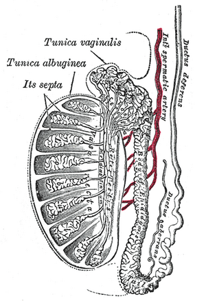 Vertical section of the testis, to show the arrangement of the ducts | |
| Details | |
| Precursor | Wolffian duct |
| Artery | Superior vesical artery, artery of the ductus deferens |
| Lymph | External iliac lymph nodes, internal iliac lymph nodes |
| Identifiers | |
| Latin | Vas deferens (plural: vasa deferentia), Ductus deferens (plural: ductus deferentes) |
| MeSH | D014649 |
| TA98 | A09.3.05.001 |
| TA2 | 3621 |
| FMA | 19234 |
| Anatomical terminology | |

The vas deferens, or ductus deferens, is part of the male reproductive system of many vertebrate. The ducts transport sperm from the epididymis to the ejaculatory ducts in anticipation of ejaculation. The vas deferens is a partially coiled tube which exits the abdominal cavity through the inguinal canal.
Etymology[]
Vas deferens is Latin, meaning "carrying-away vessel"; the plural version is vasa deferentia. Ductus deferens is also Latin, meaning "carrying-away duct"; the plural version is ductus deferentes.
Structure[]
There are two vasa deferentia, connecting the left and right epididymis with the seminal vesicles to form the ejaculatory duct in order to move sperm. In humans, each tube is about 30 centimeters (1 ft) long, 3 to 5 mm (0.118 to 0.197 inches) in diameter and is muscular (surrounded by smooth muscle). Its epithelium is pseudostratified columnar epithelium lined by stereocilia.
They are part of the spermatic cords.[1]
Blood supply[]
The vas deferens is supplied by an accompanying artery (artery of vas deferens). This artery normally arises from the superior (sometimes inferior) vesical artery, a branch of the internal iliac artery.
Nerve supply[]
The vas deferens is innervated by a variety of different types of nerve ending.[2] Adrenergic synapses are found in the smooth muscle layers.[2] Cholinergic synapses and vasoactive intestinal peptide synapses are found in the connective tissue of the mucosa.[2] Noradrenergic synapses may also be present in the vas deferens.[3]
Function[]
During ejaculation, the smooth muscle in the walls of the vas deferens contracts reflexively, thus propelling the sperm forward. This is also known as peristalsis.[4] The sperm is transferred from each vas deferens into the urethra, partially mixing with secretions from the male accessory sex glands such as the seminal vesicles, prostate gland and the bulbourethral glands, which form the bulk of semen.
Clinical significance[]
Contraception[]
A vasectomy is a method of contraception in which the vasa deferentia are permanently cut, though in some cases it can be reversed. A modern variation, which is also known as a vasectomy even though it does not include cutting the vas, involves injecting an obstructive material into the ductus to block the flow of sperm.
Investigational attempts for male contraception have focused on the vas with the use of the intra vas device and reversible inhibition of sperm under guidance.
Disease[]
The vas deferens may be obstructed, or it may be completely absent in a condition known as congenital absence of the vas deferens (CAVD, a potential feature of cystic fibrosis), causing male infertility. Acquired obstructions can occur due to infections. To treat these causes of male infertility, sperm can be harvested by testicular sperm extraction (TESE), microsurgical epididymal sperm aspiration (MESA), or other methods of collecting sperm cells directly from the testicle or epididymis.
Uses in pharmacology and physiology[]
The vas deferens has a dense sympathetic innervation,[5] making it a useful system for studying sympathetic nerve function and for studying drugs that modify neurotransmission.[6]
It has been used:
- as a bioassay for the discovery of enkephalins, the endogenous opiates.[7]
- to demonstrate quantal transmission from sympathetic nerve terminals.[8]
- as the first direct measure of free Ca2+ concentration in a postganglionic nerve terminal.[9]
- to develop an optical method for monitoring packeted transmission (similar to ).[10]
Other animals[]
Most vertebrates have some form of duct to transfer the sperm from the testes to the urethra. In cartilaginous fish and amphibians, sperm is carried through the archinephric duct, which also partially helps to transport urine from the kidneys. In teleosts, there is a distinct sperm duct, separate from the ureters, and often called the vas deferens, although probably not truly homologous with that in humans.[11] The vas deferens loops over the ureter in placental mammals, but not in marsupial mammals.[12][13]
In cartilaginous fishes, the part of the archinephric duct closest to the testis is coiled up to form an epididymis. Below this are a number of small glands secreting components of the seminal fluid. The final portion of the duct also receives ducts from the kidneys in most species.[11]
In amniotes, however, the archinephric duct has become a true vas deferens, and is used only for conducting sperm, never urine. As in cartilaginous fish, the upper part of the duct forms the epididymis. In many species, the vas deferens ends in a small sac for storing sperm.[11]
The only vertebrates to lack any structure resembling a vas deferens are the primitive jawless fishes, which release sperm directly into the body cavity, and then into the surrounding water through a simple opening in the body wall.[11]
Additional images[]

Male reproductive system.
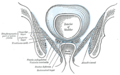
Coronal section of pelvis, showing arrangement of fasciæ. Viewed from behind.

The relations of the femoral and abdominal inguinal rings, seen from within the abdomen. Right side.

The spermatic cord in the inguinal canal.

Fundus of the bladder with the vesiculæ seminales.

Vertical section of bladder, penis, and urethra.

Prostate with seminal vesicles and seminal ducts, viewed from in front and above.

Prostate
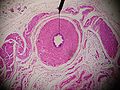
Microscopic cross section.

Testis, spermatic vessels and vas deferens

A deep dissection showing the vas deferens.


See also[]
- Intra vas device
- Excretory duct of seminal gland
- Vas deferens in the reproductive system of gastropods
References[]
- ^ Dr C Sharath Kumar, Ph D Thesis, University of Mysore, 2013
- ^ Jump up to: a b c Alm, Per (1982-07-01). "On the autonomic innervation of the human vas deferens". Brain Research Bulletin. Elsevier. 9 (1–6): 673–677. doi:10.1016/0361-9230(82)90172-1. ISSN 0361-9230. PMID 6184134. S2CID 4761228.
- ^ Mirabella, Nicola; Squillacioti, Caterina; Varricchio, Ettore; Genovese, Angelo; Paino, Giuseppe (2003-05-01). "Innervation of vas deferens and accessory male genital glands in the water buffalo (Bubalus bubalis): Neurochemical characteristics and relationships to the reproductive activity". Theriogenology. 59 (9): 1999–2016. doi:10.1016/S0093-691X(02)01260-8. ISSN 0093-691X. PMID 12600736 – via Elsevier.
- ^ Berridge, Michael J. (2008). "Smooth muscle cell calcium activation mechanisms". The Journal of Physiology. 586 (21): 5047–5061. doi:10.1113/jphysiol.2008.160440. PMC 2652144. PMID 18787034.
- ^ Sjöstrand, N.O. (1965). "The adrenergic innervation of the vas deferens and the accessory male genital organs". Acta Physiologica Scandinavica. 257: S1–82.
- ^ Burnstock, G; Verkhratsky, A (2010). "Vas deferens--a model used to establish sympathetic cotransmission". Trends in Pharmacological Sciences. 31 (3): 131–9. doi:10.1016/j.tips.2009.12.002. PMID 20074819.
- ^ Hughes, J; Smith, T. W.; Kosterlitz, H. W.; Fothergill, L. A.; Morgan, B. A.; Morris, H. R. (1975). "Identification of two related pentapeptides from the brain with potent opiate agonist activity". Nature. 258 (5536): 577–80. Bibcode:1975Natur.258..577H. doi:10.1038/258577a0. PMID 1207728. S2CID 95411.
- ^ Brock, J. A.; Cunnane, T. C. (1987). "Relationship between the nerve action potential and transmitter release from sympathetic postganglionic nerve terminals". Nature. 326 (6113): 605–7. Bibcode:1987Natur.326..605B. doi:10.1038/326605a0. PMID 2882426. S2CID 4303337.
- ^ Brain, K. L.; Bennett, M. R. (1997). "Calcium in sympathetic varicosities of mouse vas deferens during facilitation, augmentation and autoinhibition". The Journal of Physiology. 502 (3): 521–36. doi:10.1111/j.1469-7793.1997.521bj.x. PMC 1159525. PMID 9279805.
- ^ Brain, K. L.; Jackson, V. M.; Trout, S. J.; Cunnane, T. C. (2002). "Intermittent ATP release from nerve terminals elicits focal smooth muscle Ca2+ transients in mouse vas deferens". The Journal of Physiology. 541 (Pt 3): 849–62. doi:10.1113/jphysiol.2002.019612. PMC 2290369. PMID 12068045.
- ^ Jump up to: a b c d Romer, Alfred Sherwood; Parsons, Thomas S. (1977). The Vertebrate Body. Philadelphia, PA: Holt-Saunders International. pp. 393–395. ISBN 978-0-03-910284-5.
- ^ C. Hugh Tyndale-Biscoe (2005). Life of Marsupials. Csiro Publishing. ISBN 978-0-643-06257-3.
- ^ Patricia J. Armati; Chris R. Dickman; Ian D. Hume (17 August 2006). Marsupials. Cambridge University Press. ISBN 978-1-139-45742-2.
External links[]
| Wikimedia Commons has media related to Vas deferens. |
- Anatomy photo:36:07-0301 at the SUNY Downstate Medical Center—"Inguinal Region, Scrotum and Testes: Layers of the Spermatic Cord"
- Anatomy photo:44:02-0301 at the SUNY Downstate Medical Center—"The Male Pelvis: Distribution of the Peritoneum in the Male Pelvis"
- MedicalMnemonics.com: 2424 319 [dead link]
- Cross section image: pelvis/pelvis-e12-15—Plastination Laboratory at the Medical University of Vienna
- inguinalregion at The Anatomy Lesson by Wesley Norman (Georgetown University) (testes)
| show Authority control |
|---|
- Mammal male reproductive system
- Scrotum