Mediastinum testis
| Mediastinum testis | |
|---|---|
 A diagram of the major components of an adult human testicle, including the following numbered items: 1. Tunica albuginea, 2. Septula testis, 3. Lobulus testis, 4. Mediastinum testis, 5. Tubuli seminiferi contorti, 6. Tubuli seminiferi recti, 7. Rete testis, 8. Ductuli efferentes testis, 9a. Head of epididymis, 9b. Body of epididymis, 9.c Tail of epididymis,10. Vas deferens, 11a. Tunica vaginalis (parietal lamina), 11b. Tunica vaginalis (visceral lamina), and 12. Cavity of tunica vaginalis. | |
 Section of a testicle of a steer, blood vessels injected with red gelatine. 1 parenchyma, 2 mediastinum testis, 3 tunica albuginea, 4 tail of epididymis, 5 head of epididymis, 6 spermatic cord with convoluted testicular artery | |
| Identifiers | |
| TA98 | A09.3.01.018 |
| TA2 | 3595 |
| Anatomical terminology | |
The mediastinum testis is a network of fibrous connective tissue that extends from the top to near the bottom of each testis. It is wider above than below.
Numerous imperfect septa are given off from its front and sides, which radiate toward the surface of the testes and are attached to the tunica albuginea. These divide the interior of the testes into a number of incomplete spaces called lobules. These are somewhat cone-shaped, being broad at their bases at the surface of the gland, and becoming narrower as they converge to the mediastinum.
The mediastinum supports the rete testis and blood vessels of the testis in their passage to and from the substance of the gland.
Additional images[]

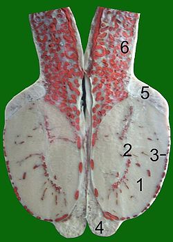
Transverse section through the left side of the scrotum and the left testis.
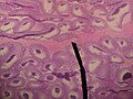
Microscopic picture.


References[]
![]() This article incorporates text in the public domain from page 1243 of the 20th edition of Gray's Anatomy (1918)
This article incorporates text in the public domain from page 1243 of the 20th edition of Gray's Anatomy (1918)
External links[]
- Histology at KUMC male-male09 "Mediastinum (human)"
- Anatomy photo:36:st-1402 at the SUNY Downstate Medical Center - "Inguinal Region, Scrotum and Testes: Testis"
| Authority control: Scientific databases |
|---|
- Wikipedia articles incorporating text from the 20th edition of Gray's Anatomy (1918)
- Mammal male reproductive system