Radial nerve
| Radial nerve | |
|---|---|
 The suprascapular, axillary, and radial nerves. | |
| Details | |
| From | posterior cord |
| To | posterior interosseous nerve |
| Innervates | posterior compartment of the arm, posterior compartment of the forearm |
| Identifiers | |
| Latin | nervus radialis |
| MeSH | D011826 |
| TA98 | A14.2.03.049 |
| TA2 | 6431 |
| FMA | 37069 |
| Anatomical terms of neuroanatomy | |
The radial nerve is a nerve in the human body that supplies the posterior portion of the upper limb. It innervates the medial and lateral heads of the triceps brachii muscle of the arm, as well as all 12 muscles in the posterior osteofascial compartment of the forearm and the associated joints and overlying skin.
It originates from the brachial plexus, carrying fibers from the ventral roots of spinal nerves C5, C6, C7, C8 & T1.[1]
The radial nerve and its branches provide motor innervation to the dorsal arm muscles (the triceps brachii and the anconeus) and the extrinsic extensors of the wrists and hands; it also provides cutaneous sensory innervation to most of the back of the hand, except for the back of the little finger and adjacent half of the ring finger (which are innervated by the ulnar nerve).
The radial nerve divides into a deep branch, which becomes the posterior interosseous nerve, and a superficial branch, which goes on to innervate the dorsum (back) of the hand.
Structure[]

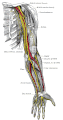
The radial nerve originates as a terminal branch of the posterior cord of the brachial plexus.[1] It goes through the arm, first in the posterior compartment of the arm, and later in the anterior compartment of the arm, and continues in the posterior compartment of the forearm.
Arm[]
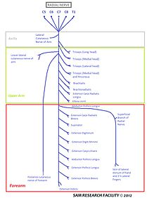
The radial nerve originates from the posterior cord of the brachial plexus with root values of C5 to C8 and T1. From the brachial plexus, it travels behind the third part of the axillary artery (part of the axillary artery distal to the pectoralis minor). In the arm, it runs behind the brachial artery and then enters the lower triangular space to reach the radial sulcus of back of the humerus.[1] It travel downwards together with profunda brachii artery, between the lateral and medial heads of triceps brachii until it reaches the lateral side the arm at 5 cm below the deltoid tuberosity where it pierces the lateral intermuscular septum to reach the anterior compartment of the arm. Then, it descends down to cross the lateral epicondyle of the humerus where the nerve terminates by branching itself into superficial and deep branch which continues into cubital fossa and then into the forearm.[2]
Radial nerve gives out muscular branches to supply the long head, medial head, and lateral head of triceps brachii muscles before and during its course in the radial sulcus. After it emerges out from the radial sulcus, it supplies the brachialis, brachioradialis and extensor carpi radialis longus.[2]
Above the radial sulcus, the radial nerve gives off posterior cutaneous nerve of the arm which supplies the skin at the back of the arm. In the radial sulcus, it gives off lower lateral cutaneous nerve of the arm and posterior cutaneous nerve of the forearm. The radial nerve also gives articular branches to supply the elbow joint.[2]
Forearm and hand[]
In the forearm, it is divided into a superficial branch (primarily sensory) and a deep branch (primarily motor).
- The superficial branch of the radial nerve is widely separated from the radial artery in the upper one third of the forearm, closely related to radial artery in the middle third of the forearm, and in the lower third, it descends in the forearm under the tendon of brachioradialis. It crosses brachioradialis to enter posterior of forearm near the back of the wrist and supply dorsum of hand. It gives sensory supply to dorsal aspect of hand, dorsal aspect of thumb, index finger, middle finger and lateral side of ring finger except the nail beds, which are supplied by proper digital branches of median nerve.[2]
- The deep branch of the radial nerve (also known as posterior interosseous nerve by some authors)[3][4]) pierces the supinator muscle, winds around the radius under the cover of supinator to reach posterior of forearm where it again pierces supinator and after which it is known as the posterior interosseous nerve. It pierces the posterior extensor muscles and comes to lie between superficial and deep muscles of the back of the forearm. At the lower border of extensor pollicis brevis, it passes deep to the extensor pollicis longus and then run on the posterior interosseous membrane. It continues to move along with posterior interosseous artery(a deep branch of common interosseous artery which is a branch of ulnar artery), and ends as a pseudoganglion below extensor retinaculum by supplying the wrist and intercarpal joints.[2]
Variation[]
It is commonly believed that the radial nerve also provided motor innervation to the long head of the triceps. However, a study conducted in 2004 found out that axillary nerve innervates the long head of triceps in 20 cadavers without any supply from radial nerve.[5]
Function[]
The following are branches of the radial nerve (including the superficial branch of the radial nerve and the deep branch of the radial nerve/posterior interosseous nerve).
Cutaneous[]

Cutaneous innervation by the radial nerve is provided by the following nerve branches:
- Posterior cutaneous nerve of arm (originates in axilla)
- Inferior lateral cutaneous nerve of arm (originates in arm)
- Posterior cutaneous nerve of forearm (originates in arm)
The superficial branch of the radial nerve provides sensory innervation to much of the back of the hand, including the web of skin between the thumb and index finger.
Motor[]

Muscular branches of the radial nerve:
- Triceps brachii
- Anconeus
- Brachioradialis
- Extensor carpi radialis longus
Deep branch of the radial nerve:
- Extensor carpi radialis brevis
- Supinator
Posterior interosseous nerve (a continuation of the deep branch after the supinator):
- Extensor digitorum
- Extensor digiti minimi
- Extensor carpi ulnaris
- Abductor pollicis longus
- Extensor pollicis brevis
- Extensor pollicis longus
- Extensor indicis
The radial nerve (and its deep branch) provides motor innervation to the muscles in the posterior compartment of the arm and forearm, which are mostly extensors.
Clinical significance[]
Injury[]
Injury to the radial nerve at different levels causes different syndromes with varying motor and sensory deficits.
At the axilla
- Common mechanisms of injury: Saturday night palsy,[1] crutch palsy, lesions[6]
- Motor deficit:
- Loss of extension of forearm, weakness of supination, and loss of extension of hand and fingers.
- Presence of wrist drop, due to inability to extend the hand and fingers.[7][8]
- Sensory deficit: Loss of sensation[7] in lateral arm, posterior forearm, the radial half of dorsum of hand, and dorsal aspect of radial 3+1⁄2 digits, excluding their nail beds.
At mid-arm
- Common mechanism of injury: Mid-shaft humeral fracture
- Motor deficit:
- Weakness of supination, and loss of extension of hand and fingers.
- Presence of wrist drop, due to inability to extend the hand and fingers.[7][8]
- Sensory deficit: Loss of sensation in posterior forearm, the radial half of dorsum of hand, and dorsal aspect of radial 3+1⁄2 digits, excluding their nail beds.
Just below the elbow
- Common mechanism of injury: Neck of radius fracture, elbow dislocation or fracture, tight cast, rheumatoid nodules, injections due to tennis elbow, injuring the deep branch of the radial nerve that pierces the radial head, causing posterior interosseous nerve syndrome
- Motor deficit:
- Weakness in extension of hand and loss of extension of fingers.
- Presence of finger drop, and partial wrist drop, since the extensor carpi radialis longus and brachioradialis muscles are working.
- Sensory deficit: None, as sensation is supplied by the superficial radial nerve
 Radial nerve

Within the distal forearm:
- Common mechanism of injury: Wartenberg's syndrome, (not to be confused with Wartenberg's sign), due to nerve entrapment beneath the tendinous insertion of brachioradialis, tight jewellery, and watch bands.
- Motor deficit: None
- Sensory deficit: Numbness and tingling in radial half of dorsum of hand, and dorsal aspect of radial 3+1⁄2 digits, excluding their nail beds.
- In Wartenberg's syndrome, there is significant radial wrist pain, and close resemblance to symptoms in de Quervain's tenosynovitis. Finkelstein's test may be positive.[9]
History[]
This section needs expansion. You can help by . (February 2014) |
Additional images[]
This gallery of anatomic features needs cleanup to abide by the medical manual of style. |

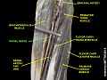
Cross-section through the middle of upper arm.

Cross-section through the middle of the forearm.

The brachial artery.

Cutaneous nerves of right upper extremity.

Superficial palmar nerves.
Nerves of the left upper extremity.

Deep palmar nerves.

Front of right upper extremity, showing surface markings for bones, arteries, and nerves.

Back of right upper extremity, showing surface markings for bones and nerves.
Radial nerve at newborn

Radial nerve
Radial nerve

Radial nerve
Radial nerve
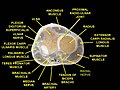
Muscles of upper limb.Cross section.


See also[]
- Muscular branches of the radial nerve
- Posterior brachial cutaneous nerve
- Dorsal antibrachial cutaneous nerve
- Superficial branch of the radial nerve
- Deep branch of the radial nerve
- Radial neuropathy
- Radial tunnel syndrome
References[]
- ^ Jump up to: a b c d Scott, Kevin R.; Ahmed, Aiesha; Scott, Linda; Kothari, Milind J. (2013-01-01), Barnes, Michael P.; Good, David C. (eds.), "Chapter 42 - Rehabilitation of brachial plexus and peripheral nerve disorders", Handbook of Clinical Neurology, Neurological Rehabilitation, Elsevier, 110: 499–514, doi:10.1016/b978-0-444-52901-5.00042-3, PMID 23312667, retrieved 2020-10-25
- ^ Jump up to: a b c d e Krishna, Garg (2010). "8 - Arm". BD Chaurasia's Human Anatomy (Regional and Applied Dissection and Clinical) Volume 1 - Upper limb and thorax (5th ed.). India: CBS Publishers and Distributors Pvt Ltd. pp. 95, 111, 122, 128. ISBN 978-81-239-1863-1.
- ^ "Acland's video Atlas of Human Anatomy 1.3.1.8 - Radial nerve in the forearm and hand". Wolters Kluwer. Archived from the original on 29 November 2017. Retrieved 26 January 2018.
The deep branch of the radial nerve, also known as the posterior interosseous nerve, is a motor nerve.
- ^ Nurul Huda, Mohd Nor; San, Aye Aye; Fauziah, Othman (2014). "An Anomalous Pattern of Superficial Branch of Radial Nerve: A Cadaveric Case Report". International Journal of Morphology. 32 (1): 29–31. doi:10.4067/S0717-95022014000100005. ISSN 0717-9502.
The deep branch of radial nerve and also known as posterior interosseous nerve is the nerve of extensor compartment of the forearm ...
- ^ de Sèze MP, Rezzouk J, de Sèze M, Uzel M, Lavignolle B, Midy D, Durandeau A (2004). "Does the motor branch of the long head of the triceps brachii arise from the radial nerve? An anatomic and electromyographic study". Surg Radiol Anat. 26 (6): 459–61. doi:10.1007/s00276-004-0253-z. PMID 15365769.
- ^ Ryan, Monique M.; Jones, H. Royden (2015-01-01), Darras, Basil T.; Jones, H. Royden; Ryan, Monique M.; De Vivo, Darryl C. (eds.), "Chapter 14 - Mononeuropathies", Neuromuscular Disorders of Infancy, Childhood, and Adolescence (Second Edition), San Diego: Academic Press, pp. 243–273, doi:10.1016/b978-0-12-417044-5.00014-7, ISBN 978-0-12-417044-5, retrieved 2020-10-25
- ^ Jump up to: a b c Prakash, M. V. S. Satya; Udupi Bidkar, Prasanna (2016-01-01), Prabhakar, Hemanshu (ed.), "Chapter 37 - Peripheral Nerve Injuries", Complications in Neuroanesthesia, San Diego: Academic Press, pp. 359–368, doi:10.1016/b978-0-12-804075-1.00037-7, ISBN 978-0-12-804075-1, retrieved 2020-10-25
- ^ Jump up to: a b Kretschmer, Thomas; Heinen, Christian (2015-01-01), Tubbs, R. Shane; Rizk, Elias; Shoja, Mohammadali M.; Loukas, Marios (eds.), "Chapter 36 - Iatrogenic Injuries of the Nerves", Nerves and Nerve Injuries, San Diego: Academic Press, pp. 557–585, doi:10.1016/b978-0-12-802653-3.00085-3, ISBN 978-0-12-802653-3, retrieved 2020-10-25
- ^ Tor Wo Chiu (2011). Stone's Plastic Surgery Facts and Figures. Cambridge University Press. ISBN 9781139499781.
External links[]
| Wikimedia Commons has media related to Radial nerve. |
- Radial_nerve at the Duke University Health System's Orthopedics program
- Atlas image: hand_plexus at the University of Michigan Health System - "Axilla, dissection, anterior view"
| show Authority control |
|---|
- Nerves of the upper limb