Skeleton
This article needs additional citations for verification. (August 2021) |
| Skeleton | |
|---|---|
 | |
| Details | |
| Identifiers | |
| Greek | σκελετός |
| MeSH | D012863 |
| TA98 | A02.0.00.000 |
| TA2 | 352 |
| FMA | 2021 |
| Anatomical terminology | |
A skeleton is a structural frame that supports an animal body.[1] There are several different skeletal types: the exoskeleton, which is the stable outer shell of an organism, the endoskeleton, which forms the support structure inside the body, the hydroskeleton, a flexible skeleton supported by fluid pressure, and the cytoskeleton present in the cytoplasm of all cells, including bacteria, and archaea.[clarification needed] The term comes from Greek σκελετός (skeletós) 'dried up'.[2]
Types of skeletons[]
There are two major types of skeletons: solid and fluid. Solid skeletons can be internal, called an endoskeleton, or external, called an exoskeleton, and may be further classified as pliant (elastic/movable) or rigid (hard/non-movable).[3] Fluid skeletons are always internal.
Exoskeleton[]

Exoskeletons are external, and are found in many invertebrates; they enclose and protect the soft tissues and organs of the body. Some kinds of exoskeletons undergo periodic moulting or ecdysis as the animal grows, as is the case in many arthropods including insects and crustaceans.
The exoskeleton of insects is not only a form of protection, but also serves as a surface for muscle attachment, as a watertight protection against drying, and as a sense organ to interact with the environment. The shell of mollusks also performs all of the same functions, except that in most cases it does not contain sense organs.
An external skeleton can be quite heavy in relation to the overall mass of an animal, so on land, organisms that have an exoskeleton are mostly relatively small. Somewhat larger aquatic animals can support an exoskeleton because weight is less of a consideration underwater. The southern giant clam, a species of extremely large saltwater clam in the Pacific Ocean, has a shell that is massive in both size and weight. Syrinx aruanus is a species of sea snail with a very large shell.
Endoskeleton[]

The endoskeleton is the internal support structure of an animal, composed of mineralized tissue and is typical of vertebrates. Endoskeletons vary in complexity from functioning purely for support (as in the case of sponges), to serving as an attachment site for muscles and a mechanism for transmitting muscular forces. A true endoskeleton is derived from mesodermal tissue. Such a skeleton is present in echinoderms and chordates.
Pliant skeletons[]
Pliant skeletons are capable of movement; thus, when stress is applied to the skeletal structure, it deforms and then reverts to its original shape. This skeletal structure is used in some invertebrates, for instance in the hinge of bivalve shells or the mesoglea of cnidarians such as jellyfish. Pliant skeletons are beneficial because only muscle contractions are needed to bend the skeleton; upon muscle relaxation, the skeleton will return to its original shape. Cartilage is one material that a pliant skeleton may be composed of, but most pliant skeletons are formed from a mixture of proteins, polysaccharides, and water.[3] For additional structure or protection, pliant skeletons may be supported by rigid skeletons. Organisms that have pliant skeletons typically live in water, which supports body structure in the absence of a rigid skeleton.[4]
Rigid skeletons[]
Rigid skeletons are not capable of movement when stressed, creating a strong support system most common in terrestrial animals. Such a skeleton type used by animals that live in water are more for protection (such as barnacle and snail shells) or for fast-moving animals that require additional support of musculature needed for swimming through water. Rigid skeletons are formed from materials including chitin (in arthropods), calcium compounds such as calcium carbonate (in stony corals and mollusks) and silicate (for diatoms and radiolarians).
Cytoskeleton[]
The cytoskeleton (cyto- meaning cell[5]) is used to stabilize and preserve the form of the cells. It is a dynamic structure that maintains cell shape, protects the cell, enables cellular motion (using structures such as flagella, cilia and lamellipodia), and plays important roles in both intracellular transport (the movement of vesicles and organelles, for example) and cellular division.
Fluid skeletons[]
Hydrostatic skeleton (hydroskeleton)[]
A hydrostatic skeleton is a semi-rigid, soft tissue structure filled with liquid under pressure, surrounded by muscles. Longitudinal and circular muscles around their body sectors allow movement by alternate lengthening and contractions along their lengths. A common example of this is the earthworm.
Organisms with skeletons[]
Invertebrates[]
The endoskeletons of echinoderms and some other soft-bodied invertebrates such as jellyfish and earthworms are also termed hydrostatic; a body cavity the coelom is filled with coelomic fluid and the pressure from this fluid acts together with the surrounding muscles to change the organism's shape and produce movement.
Sponges[]
The skeleton of sponges consists of microscopic calcareous or silicious spicules. The demosponges include 90% of all species of sponges. Their "skeletons" are made of spicules consisting of fibers of the protein spongin, the mineral silica, or both. Where spicules of silica are present, they have a different shape from those in the otherwise similar glass sponges.[6]
Echinoderms[]
The skeleton of the echinoderms, which include, among other things, the starfish, is composed of calcite and a small amount of magnesium oxide. It lies below the epidermis in the mesoderm and is within cell clusters of frame-forming cells. This structure formed is porous and therefore firm and at the same time light. It coalesces into small calcareous ossicles (bony plates), which can grow in all directions and thus can replace the loss of a body part. Connected by joints, the individual skeletal parts can be moved by the muscles.
Vertebrates[]

In most vertebrates, the main skeletal component is referred to as bone. These bones compose a unique skeletal system for each type of animal. Another important component is cartilage which in mammals is found mainly in the joint areas. In other animals, such as the cartilaginous fishes, which include the sharks, the skeleton is composed entirely of cartilage. The segmental pattern of the skeleton is present in all vertebrates (mammals, birds, fish, reptiles and amphibians) with basic units being repeated. This segmental pattern is particularly evident in the vertebral column and the ribcage.
Bones in addition to supporting the body also serve, at the cellular level, as calcium and phosphate storage.
Fish[]
The skeleton, which forms the support structure inside the fish is either made of cartilage as in the (Chondrichthyes), or bones as in the (Osteichthyes). The main skeletal element is the vertebral column, composed of articulating vertebrae which are lightweight yet strong. The ribs attach to the spine and there are no limbs or limb girdles. They are supported only by the muscles. The main external features of the fish, the fins, are composed of either bony or soft spines called rays, which with the exception of the caudal fin (tail fin), have no direct connection with the spine. They are supported by the muscles which compose the main part of the trunk.
Birds[]
The bird skeleton is highly adapted for flight. It is extremely lightweight, yet still strong enough to withstand the stresses of taking off, flying, and landing. One key adaptation is the fusing of bones into single ossifications, such as the pygostyle. Because of this, birds usually have a smaller number of bones than other terrestrial vertebrates. Birds also lack teeth or even a true jaw, instead having evolved a beak, which is far more lightweight. The beaks of many baby birds have a projection called an egg tooth, which facilitates their exit from the amniotic egg.
Marine mammals[]
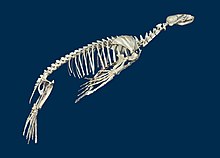
To facilitate the movement of marine mammals in water, the hind legs were either lost altogether, as in the whales and manatees, or united in a single tail fin as in the pinnipeds (seals). In the whale, the cervical vertebrae are typically fused, an adaptation trading flexibility for stability during swimming.[citation needed]
Humans[]

The skeleton consists of both fused and individual bones supported and supplemented by ligaments, tendons, muscles and cartilage. It serves as a scaffold which supports organs, anchors muscles, and protects organs such as the brain, lungs, heart and spinal cord. Although the teeth do not consist of tissue commonly found in bones, the teeth are usually considered as members of the skeletal system.[7] The biggest bone in the body is the femur in the upper leg, and the smallest is the stapes bone in the middle ear. In an adult, the skeleton comprises around 13.1% of the total body weight,[8] and half of this weight is water.
Fused bones include those of the pelvis and the cranium. Not all bones are interconnected directly: There are three bones in each middle ear called the ossicles that articulate only with each other. The hyoid bone, which is located in the neck and serves as the point of attachment for the tongue, does not articulate with any other bones in the body, being supported by muscles and ligaments.
There are 206 bones in the adult human skeleton, although this number depends on whether the pelvic bones (the hip bones on each side) are counted as one or three bones on each side (ilium, ischium, and pubis), whether the coccyx or tail bone is counted as one or four separate bones, and does not count the variable wormian bones between skull sutures. Similarly, the sacrum is usually counted as a single bone, rather than five fused vertebrae. There is also a variable number of small sesamoid bones, commonly found in tendons. The patella or kneecap on each side is an example of a larger sesamoid bone. The patellae are counted in the total, as they are constant. The number of bones varies between individuals and with age – newborn babies have over 270 bones some of which fuse together.[citation needed] These bones are organized into a longitudinal axis, the axial skeleton, to which the appendicular skeleton is attached.[9]
The human skeleton takes 20 years before it is fully developed, and the bones contain marrow, which produces blood cells.
There exist several general differences between the male and female skeletons. The male skeleton, for example, is generally larger and heavier than the female skeleton. In the female skeleton, the bones of the skull are generally less angular. The female skeleton also has wider and shorter breastbone and slimmer wrists. There exist significant differences between the male and female pelvis which are related to the female's pregnancy and childbirth capabilities. The female pelvis is wider and shallower than the male pelvis. Female pelvises also have an enlarged pelvic outlet and a wider and more circular pelvic inlet. The angle between the pubic bones is known to be sharper in males, which results in a more circular, narrower, and near heart-shaped pelvis.[10][11]
Parts[]
Bone[]
Bones are rigid organs that form part of the endoskeleton of vertebrates. They function to move, support, and protect the various organs of the body, produce red and white blood cells and store minerals. Bone tissue is a type of dense connective tissue. Bones have a variety of shapes with a complex internal and external structure they are also lightweight, yet strong and hard. One of the types of tissue that makes up bone tissue is mineralized tissue and this gives it rigidity and a honeycomb-like three-dimensional internal structure. Other types of tissue found in bones include marrow, endosteum and periosteum, nerves, blood vessels and cartilage.
Extra-skeletal bones in mammals[]
These bones, primarily formed separately in subcutaneous tissues, include headgears (such as bony core of horns, antlers, and ossicones), osteoderm, and os penis/ os clitoris.[12]
Cartilage[]
During embryonic development the precursor to bone development is cartilage that mostly becomes replaced by bone, after flesh such as muscle has formed around it. Cartilage is a stiff and inflexible connective tissue found in many areas including the joints between bones, the rib cage, the ear, the nose, the elbow, the knee, the ankle, the bronchial tubes and the intervertebral discs. It is not as hard and rigid as bone but is stiffer and less flexible than muscle.
Cartilage is composed of specialized cells called chondrocytes that produce a large amount of extracellular matrix composed of Type II collagen (except fibrocartilage which also contains type I collagen) fibers, abundant ground substance rich in proteoglycans, and elastin fibers. Cartilage is classified in three types, elastic cartilage, hyaline cartilage and fibrocartilage, which differ in the relative amounts of these three main components.
Unlike other connective tissues, cartilage does not contain blood vessels. The chondrocytes are supplied by diffusion, helped by the pumping action generated by compression of the articular cartilage or flexion of the elastic cartilage. Thus, compared to other connective tissues, cartilage grows and repairs more slowly.
Ligament[]
A ligament is a piece of rubbery tissue that connects bone to other bone.[13] It is commonly confused with the tendon, a similar structure that connects muscle to bone.
Tendon[]
A tendon is a rubber-band like tissue that connects muscle to bone. It is not to be confused with the ligament, a similar tissue that connects bone to bone.
See also[]
References[]
- ^ Hamilton, William James; Manton, Sidnie M. (2001). "Skeleton". Encyclopedia Britannica (online ed.). Retrieved 25 December 2020.
- ^ "skeleton". Mish 2014, p. 1167.
- ^ Jump up to: a b Ruppert, Fox & Barnes 2003, p. 102.
- ^ Pechenik 2015.[page needed]
- ^ "cyt- or cyto-". Mish 2014, p. 312.
- ^ Barnes, Fox & Barnes, pp. 105–6.
- ^ "Skeletal System: Facts, Function & Diseases". Live Science. Archived from the original on 7 March 2017. Retrieved 7 March 2017.
- ^ Reynolds & Karlotski 1977, p. 161
- ^ Tözeren 2000, pp. 6–10.
- ^ Balaban 2008, p. 61
- ^ Stein 2007, p. 73.
- ^ "Formation, structure, and function of extra‐skeletal bones in mammals" (PDF). Biological Reviews. 2020. doi:10.1111/brv.12597.
- ^ Vorvick, Linda J. (13 August 2020). "Tendon vs. Ligament". A.D.A.M. Medical Encyclopedia. Ebix. Retrieved 6 August 2021 – via MedLinePlus.
Bibliography[]
- Balaban, Naomi (2008). The Handy Anatomy Answer Book. Visible Ink Press. ISBN 978-1-57859-190-9.
- Forbes, R. M.; Mitchell, H. H.; Cooper, A. R. (1956). "Further studies on the gross composition and mineral elements of the adult human body". Journal of Biological Chemistry. American Society for Biochemistry and Molecular Biology. 223 (2): 969–75. doi:10.1016/S0021-9258(18)65095-1. PMID 13385244.
- Mish, Frederick C., ed. (2003). Merriam-Webster's Collegiate Dictionary (11th ed.). Merriam-Webster. ISBN 978-0-87779-807-1.
- Nasoori, Alireza (2020). "Formation, structure, and function of extra‐skeletal bones in mammals" (PDF). Biological Reviews. Cambridge Philosophical Society. 95 (4): 986–1019. doi:10.1111/brv.12597. PMID 32338826. S2CID 216556342.
- Pechenik, Jan A. (2015). Biology of the Invertebrates (7th ed.). McGraw-Hill Education. ISBN 978-0-07-352418-4.
- Reynolds, William W.; Karlotski, William J. (1977). "The Allometric Relationship of Skeleton Weight to Body Weight in Teleost Fishes: A Preliminary Comparison with Birds and Mammals". Copeia. American Society of Ichthyologists and Herpetologists. 1977 (1): 160–3. doi:10.2307/1443520. JSTOR 1443520.
- Ruppert, Edward E.; Fox, Richard S.; Barnes, Robert D. (2003). Invertebrate Zoology (7th ed.). Thomson, Brooks/Cole. ISBN 978-0-03-025982-1.
- Stein, Lisa (2007). Body: The Complete Human. National Geographic Society. ISBN 978-1-4262-0128-8.
- Tözeren, Aydın (2000). Human Body Dynamics: Classical Mechanics and Human Movement. Springer. ISBN 0-387-98801-7.
External links[]
![]() Media related to Skeletons at Wikimedia Commons
Media related to Skeletons at Wikimedia Commons
| The Wikibook Anatomy and Physiology of Animals has a page on the topic of: The Skeleton |
- 3-D Viewer of a male American mastodon skeleton, with bones labelled, at the University of Michigan Mammutidae digital fossil repository
- Interactive views of various primate skeletons at eSkeletons.org (associated with the University of Texas at Austin
| hide Authority control | |
|---|---|
| General | |
| National libraries | |
| Scientific databases | |
- Skeletons
- Animal anatomy