Theca of follicle
This article's factual accuracy is disputed. (May 2015) |
| Theca of follicle | |
|---|---|
| Details | |
| Identifiers | |
| Latin | theca folliculi |
| MeSH | D013799 |
| Anatomical terminology | |
The theca folliculi comprise a layer of the ovarian follicles. They appear as the follicles become secondary follicles.
The theca are divided into two layers, the theca interna and the theca externa.[citation needed]
Theca cells are a group of endocrine cells in the ovary made up of connective tissue surrounding the follicle. They have many diverse functions, including folliculogenesis.[1]
Androgen synthesis[]

Theca cells are responsible for synthesizing androgens, providing signal transduction between granulosa cells and oocytes during development by the establishment of a vascular system, providing nutrients, and providing structure and support to the follicle as it matures.[1]
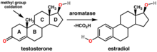
Theca cells are responsible for the production of androstenedione, and indirectly the production of 17β estradiol, also called E2, by supplying the neighboring granulosa cells with androstenedione that with the help of the enzyme aromatase can be used as a substrate for this type of estradiol.[2] FSH induces the granulosa cells to make aromatase that converts the androgens made by the theca interna into estradiol.[3]
Signaling cascade[]
Theca cells are stimulated by luteinizing hormone (LH), with signaling originating in the hypothalamus of the brain.
Gonadotropin releasing hormone (GnRH) is released by projections of the hypothalamus into the anterior pituitary gland. Gonadotrophs are stimulated to produce follicle-stimulating hormone (FSH) and luteinizing hormone (LH), which are released into the bloodstream to act upon the ovaries.
Within the ovaries, transmembrane G-protein coupled receptors (GPCRs) bind to LH in the bloodstream, and the signal is transduced to the interior of theca cells through the action of the second messenger cAMP and third messenger protein kinase A (PKA). Theca cells are then stimulated to produce testosterone, which is sent in a paracrine fashion to neighboring granulosa cells for conversion to estradiol.[4]
Disorders[]
Hyperactivity of theca cells causes hyperandrogenism, and hypoactivity leads to a lack of estrogen.[5] Granulosa cell tumors, while rare (less than 5% of ovarian cancers), involve both the granulosa cells and the theca cells.[6]
Folliculogenesis[]

In human adult females, the primordial follicle is composed of a single oocyte surrounded by a layer of closely associated granulosa cells. In early stages of the ovarian cycle, the developing follicle acquires a layer of connective tissue and associated blood vessels. This covering is called the theca.
As development of the secondary follicle progresses, granulosa cells proliferate to form the multilayered membrana granulosum. Over a period of months, the granulosa cells and thecal cells secrete antral fluid (a mixture of hormones, enzymes, and anticoagulants) to nourish the maturing ovum.
In tertiary follicles, the single-layered theca differentiates into a theca interna and theca externa. The theca interna contains glandular cells and many small blood vessels, while the theca externa is composed of dense connective tissue and larger blood vessels.[7]
Structural roles[]
Theca cells (along with granulosa cells) form the corpus luteum during oocyte maturation. Theca cells are only correlated with developing ovarian follicles.[5] They are the leading cause of endocrine-based infertility, as either hyperactivity or hypoactivity of the theca cells can lead to fertility problems.
See also[]
- ovary
- theca
- thecoma
- polycystic ovarian syndrome
- dehydroepiandrosterone sulfate
- luteinizing hormone
- follicle-stimulating hormone
References[]
- ^ Jump up to: a b Young, J. M.; McNeilly, A. S. (2010). "Theca: the forgotten cell of the ovarian follicle". Reproduction. 140 (4): 489–504. doi:10.1530/REP-10-0094. PMID 20628033.
- ^ Hall, John E. (John Edward), 1946- (2015-05-20). Guyton and Hall textbook of medical physiology (13th ed.). Philadelphia, PA. p. 1042. ISBN 9781455770052. OCLC 900869748.CS1 maint: multiple names: authors list (link)
- ^ Hall, John E. (John Edward), 1946- (2015-05-20). Guyton and Hall textbook of medical physiology (13th ed.). Philadelphia, PA. p. 1044. ISBN 9781455770052. OCLC 900869748.CS1 maint: multiple names: authors list (link)
- ^ Medical physiology. Boron, Walter F.,, Boulpaep, Emile L. (Third ed.). Philadelphia, PA. 2016-03-29. ISBN 978-1-4557-3328-6. OCLC 951680737.CS1 maint: others (link)
- ^ Jump up to: a b Magoffin, Denis A. (2005). "Ovarian theca cell". The International Journal of Biochemistry & Cell Biology. 37 (7): 1344–9. doi:10.1016/j.biocel.2005.01.016. PMID 15833266.
- ^ Kottarathil, Vijaykumar Dehannathparambil; Antony, Michelle Aline; Nair, Indu R.; Pavithran, Keechilat (2013). "Recent Advances in Granulosa Cell Tumor Ovary: A Review". Indian Journal of Surgical Oncology. 4 (1): 37–47. doi:10.1007/s13193-012-0201-z. PMC 3578540. PMID 24426698.
- ^ Jones, Richard E. (Richard Evan), 1940- (2006). Human reproductive biology. Lopez, Kristin H. (3rd ed.). Amsterdam: Elsevier Academic Press. ISBN 978-0-12-088465-0. OCLC 61351645.CS1 maint: multiple names: authors list (link)
External links[]
- Histology image: 14805loa – Histology Learning System at Boston University
- Anatomy photo: Reproductive/mammal/ovary2/ovary5 - Comparative Organology at University of California, Davis - "Mammal, canine ovary (LM, High)"
- Anatomy photo: Reproductive/mammal/ovary5/ovary6 - Comparative Organology at University of California, Davis - "Mammal, bovine ovary (LM, Medium)"
- UIUC Histology Subject 372 - interna
- UIUC Histology Subject 373 - externa
- Anatomy Atlases - Microscopic Anatomy, plate 13.249
- Slide at trinity.edu
This women's health related article is a stub. You can help Wikipedia by . |
This article related to the genitourinary system is a stub. You can help Wikipedia by . |
- Mammal female reproductive system
- Steroid hormone secreting cells
- Women's health stubs
- Genitourinary system stubs