Abdominopelvic cavity
| Abdominopelvic cavity | |
|---|---|
 | |
| Details | |
| Identifiers | |
| Latin | cavitas abdominis et pelvis |
| TA98 | A01.1.00.050 A10.1.00.000 |
| TA2 | 3699 |
| FMA | 12267 |
| Anatomical terminology | |
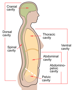
The abdominopelvic cavity is a body cavity that consists of the abdominal cavity and the pelvic cavity.[1] It contains the stomach, liver, pancreas, spleen, gallbladder, kidneys, and most of the small and large intestines. It also contains the urinary bladder and internal reproductive organs. The abdominal pelvic cavity is a little pocket sac that lies way low in the base of the abdominal pelvis cavity. There's no membrane that separates out the abdominal cavity from the pelvic cavity so it is sometimes referred to as the abdominal pelvis or the peritoneal cavity. There are many diseases and disorders associated with the organs of the abdominopelvic cavity.
The stomach sits on the left side, which is attached to the esophagus tube. Food comes through the esophagus, goes behind all of the other organs in the thoracic cavity then comes out through the esophagus and opens up into the stomach. The stomach is more of a mashing and acidic environment which begins the major processes of digestion. The food particles must be broken down before entering the small intestine. The stomach can be affected by a few types of diseases. Helicobacter pylorus, previously known as Campylobacter pylori is a Gram-negative, microaerophilic bacterium infection usually found in the stomach. The article called “Gastric Campylobacter-like organisms, gastritis, and peptic ulcer disease” states that Campylobacter pylori infection is “known to be the most common and important cause of gastritis, and C. pylori infections have been associated with duodenal ulcer, gastric ulcer, nonulcer dyspepsia, and gastric cancer” (Graham 1). I know one or two people who have the infection. People who are infected with the Campylobacter pylori should drink extra fluids as long as the diarrhea lasts. Antibiotics are needed or prescribed to people who are very ill or at high risk for severe disease, such as people with severely weakened immune systems. For example, people with the blood disorders thalassemia and hypogammaglobulinemia, AIDS, or people receiving chemotherapy.
The small intestine is about 20 feet and goes behind the big large intestine then makes a mass of curly tube. The small intestine is divided into 3 parts: duodenum, jejunum and ileum. The Duodenum receives particles from different organs like, the pancreas. The pancreas will start secreting things into the first section of the duodenum. The second part is the jejunum and it is located in the middle of the small intestine. When looking at a model, one cannot tell when it stops and starts, therefore, you'll need to look as logically as possible to try and figure out exactly where it is. Then the final part of the small intestine is the ilium. The ilium is connected right into the large intestine. The ileum is connected right into the ileum cecum valve which is the beginning of the large intestine. The large intestine, also known as the large bowel, is the last part of the gastrointestinal tract and of the digestive system. One disease that affects the lining of the GI tract. Crohn’s disease is a chronic inflammatory condition of the gastrointestinal tract. Crohn's most commonly affects the end of the small bowel (the ileum) and the beginning of the colon, but it may affect any part of the gastrointestinal (GI) tract, from the mouth to the anus. The article “Crohn's disease of the large intestine” states that “ The diagnosis of Crohn’s disease can sometimes be established or confirmed by examination of tissues of removed at laparotomy when resection of the intestinal lesions is indicated” (Morson, 502). Overall, Crohn's disease affects both large and small intestine.
The pancreas produces the hormone, insulin, which helps controls your blood sugar. The spleen, on a model is a curved purple shaped organ which is located on the left side, hidden as a cavity organ. The spleen filters out your red blood cells and pulls out your old blood cells and cleans them out. The spleen also sits right against the ribs so if you crack a rib, you can puncture the spleen. There's a lot of blood flowing in the spleen, so there is a higher chance of excessive bleeding. Acute pancreatitis is sudden inflammation of the pancreas that may be mild or life-threatening but usually subsides. Gallstones and excessive alcohol use are the main causes of acute pancreatitis. Severe abdominal pain is the predominant symptom. In the article “Acute Pancreatitis” it states that “The disease occurs at a similar frequency among various age groups, but the cause of the condition and the likelihood of death vary according to age, sex, race, body-mass index (the weight in kilograms divided by the square of the height in meters), and other factors”. The article goes on to say “most important risk factors for pancreatitis in adults are gallstones and excessive alcohol use, although clinically detected pancreatitis never develops in most persons with these risk factors”. The pancreas is an organ that regulates blood sugar and pancreatitis is the inflammation of the pancreas.
Coming off the cecum (tiny tail piece) is the appendix. The appendix is a small organ attached to the large intestine in the lower right side of the belly. When it gets infected, it's called appendicitis. When there is a buildup of bacteria, it can get in flamed and swollen and that leads to appendicitis. . Appendicitis is an inflammation of the appendix. It is a finger-shaped pouch that projects from your colon on the lower right side of your abdomen. The appendix doesn't seem to have a specific purpose; the cause of it is unknown. The article “Acute appendicitis” says that “Appendicitis is the most common abdominal emergency and accounts for more than 40 000 hospital admissions in England every year. Appendicitis is most common between the ages of 10 and 20 years, but no age is exempt. A male preponderance exists, with a male to female ratio of 1.4:1; the overall lifetime risk is 8.6% for males and 6.7% for females in the United States” (Simpson 1). Although there is no function for appendix, appendicitis is the most common abdominal emergency.
The liver is a processing and detoxifying organ. It filters all the blood and cleans It out. Also when looking at a diagram, the liver sits right up against the diaphragm on the right side. The gallbladder (usually the color green on most diagrams) on the other hand is located on the inferior (below) and posterior (backside) surface of the liver. The gallbladder produces bile, which is used to help process fats in the body. Humans can live without the gallbladder. People have them remove all the time, but it is important to be extra careful with your diet and what kind of fatty things you choose to put in the body. A consequence may result in getting diarrhea, due to fact that your body is no longer processing the food as well. Cirrhosis is a complication of many liver diseases characterized by abnormal structure and function of the liver. The diseases that lead to cirrhosis do so because they injure and kill liver cells, after which the inflammation and repair that is associated with the dying liver cells causes, scar tissue to form. The liver cells that do not die multiply in an attempt to replace the cells that have died. This results in clusters of newly formed liver cells (regenerative nodules) within the scar tissue. The article Liver cirrhosis states “The exact prevalence of cirrhosis worldwide is unknown. It was estimated at 0·15% or 400 000 in the USA, 7 which accounted for more than 25000 deaths and 373000 hospital discharges in 1998”.[2] Therefore, the liver is processing and detoxifying organ but leaves to a very high rate of deaths due to cirrhosis.
On the very dorsal aspect of an abdominal cavity model, there's two little kidney bean shaped organs that are called the Left and right kidney. The kidneys filter the blood and urine. Urinary tract infections are caused by microbes such as bacteria overcoming the body's defenses in the urinary tract. They can affect the kidneys, bladder, and the tubes that run between them. The urinary tract can be divided into the upper urinary tract and the lower urinary tract. The upper urinary tract consists of the kidneys and the ureters, and the lower urinary tract consists of the bladder and the urethra. They are one of the most common types of infection and account for around 8.1 million visits to a doctor every year. In the “Bacterial urinary Tract Infections in Diabetes “states that “urinary tract is significant problem in patients with diabetes mellitus because of the multiple effects of this disease on the immune system. Complicated urinary tract infections associated with diabetes include renal and perirenal abscess, the gas –forming infections, such emphysematous pyelonephritis, and renal papillary necrosis” (Andriole 1). In conclusion, the kidney filters blood and urine but in some cases can lead to minor infections such as UTIs.
Additional images[]

The abdominopelvic cavity 6 is made up of the abdominal cavity 3 and the pelvic cavity 4.
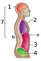
The abdominopelvic cavity is made up of the abdominal cavity 3 and the pelvic cavity 4.


References[]
- ^ "abdominopelvic cavity". NDI Foundation. Archived from the original on 20 February 2013. Retrieved 2 April 2013.
- ^ Schuppan, Detlef; Afdhal, Nezam H. (2008). "Liver cirrhosis". The Lancet. 371 (9615): 838–851. doi:10.1016/S0140-6736(08)60383-9. PMC 2271178. PMID 18328931.
- Graham, David Y. "Campylobacter pylori and peptic ulcer disease." Gastroenterology 96.2 (1989): 615–625.
- Lockhart-Mummery, H. E., and B. C. Morson. "Crohn's disease of the large intestine." Gut 5.6 (1964):
- Humes, D. J., and J. Simpson. "Acute appendicitis." Bmj 333.7567 (2006): 530–534.
- Patterson, Jan Evans, and Vincent T. Andriole. "Bacterial urinary tract infections in diabetes." Infectious Disease Clinics 11.3 (1997): 735–750.
| Authority control: Scientific databases |
|---|
- Abdomen