Pineal gland
| Pineal gland | |
|---|---|
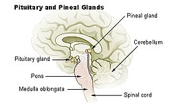 Diagram of pituitary and pineal glands in the human brain | |
| Details | |
| Precursor | Neural ectoderm, roof of diencephalon |
| Artery | Posterior cerebral artery |
| Identifiers | |
| Latin | Glandula pinealis |
| MeSH | D010870 |
| NeuroNames | 297 |
| NeuroLex ID | birnlex_1184 |
| TA98 | A11.2.00.001 |
| TA2 | 3862 |
| FMA | 62033 |
| Anatomical terms of neuroanatomy | |

The pineal gland, conarium, or epiphysis cerebri, is a small endocrine gland in the brain of most vertebrates. The pineal gland produces melatonin, a serotonin-derived hormone which modulates sleep patterns in both circadian and seasonal cycles. The shape of the gland resembles a pine cone, which gives it its name.[1] The pineal gland is located in the epithalamus, near the center of the brain, between the two hemispheres, tucked in a groove where the two halves of the thalamus join.[2][3] The pineal gland is one of the neuroendocrine secretory circumventricular organs in which capillaries are mostly permeable to solutes in the blood.[4]
Nearly all vertebrate species possess a pineal gland. The most important exception is a primitive vertebrate, the hagfish. Even in the hagfish, however, there may be a "pineal equivalent" structure in the dorsal diencephalon.[5] The lancelet Branchiostoma lanceolatum, the nearest existing relative to vertebrates, also lacks a recognizable pineal gland.[6] The lamprey (another primitive vertebrate), however, does possess one.[6] A few more complex vertebrates[which?] have lost pineal glands over the course of their evolution.[7]
The results of various scientific research in evolutionary biology, comparative neuroanatomy and neurophysiology have explained the evolutionary history (phylogeny) of the pineal gland in different vertebrate species. From the point of view of biological evolution, the pineal gland is a kind of atrophied photoreceptor. In the epithalamus of some species of amphibians and reptiles, it is linked to a light-sensing organ, known as the parietal eye, which is also called the pineal eye or third eye.[8]
René Descartes believed the human pineal gland to be the "principal seat of the soul." Academic philosophy among his contemporaries considered the pineal gland as a neuroanatomical structure without special metaphysical qualities; science studied it as one endocrine gland among many.[9]
Etymology[]
The word pineal, from Latin pinea (pine-cone), was first used in the late 17th century to refer to the cone shape of the brain gland.[1]
Structure[]
The pineal gland is a midline brain structure that is unpaired. It takes its name from its pine-cone shape.[1][10] The gland is reddish-gray and about the size of a grain of rice (5–8 mm) in humans. The pineal gland, also called the pineal body, is part of the epithalamus, and lies between the laterally positioned thalamic bodies and behind the habenular commissure. It is located in the quadrigeminal cistern near to the corpora quadrigemina.[11] It is also located behind the third ventricle and is bathed in cerebrospinal fluid supplied through a small pineal recess of the third ventricle which projects into the stalk of the gland.[12]
Blood supply[]
Unlike most of the mammalian brain, the pineal gland is not isolated from the body by the blood–brain barrier system;[13] it has profuse blood flow, second only to the kidney,[14] supplied from the choroidal branches of the posterior cerebral artery.
Nerve supply[]
The pineal gland receives a sympathetic innervation from the superior cervical ganglion. A parasympathetic innervation from the pterygopalatine and otic ganglia is also present.[15] Further, some nerve fibers penetrate into the pineal gland via the pineal stalk (central innervation). Also, neurons in the trigeminal ganglion innervate the gland with nerve fibers containing the neuropeptide PACAP.
Microanatomy[]
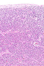
The pineal body in humans consists of a lobular parenchyma of pinealocytes surrounded by connective tissue spaces. The gland's surface is covered by a pial capsule.
The pineal gland consists mainly of pinealocytes, but four other cell types have been identified. As it is quite cellular (in relation to the cortex and white matter), it may be mistaken for a neoplasm.[16]
| Cell type | Description |
|---|---|
| Pinealocytes | The pinealocytes consist of a cell body with 4–6 processes emerging. They produce and secrete melatonin. The pinealocytes can be stained by special silver impregnation methods. Their cytoplasm is lightly basophilic. With special stains, pinealocytes exhibit lengthy, branched cytoplasmic processes that extend to the connective septa and its blood vessels. |
| Interstitial cells | Interstitial cells are located between the pinealocytes. They have elongated nuclei and a cytoplasm that is stained darker than that of the pinealocytes. |
| Perivascular phagocyte | Many capillaries are present in the gland, and perivascular phagocytes are located close to these blood vessels. The perivascular phagocytes are antigen presenting cells. |
| Pineal neurons | In higher vertebrates neurons are usually located in the pineal gland. However, this is not the case in rodents. |
| Peptidergic neuron-like cells | In some species, neuronal-like peptidergic cells are present. These cells might have a paracrine regulatory function. |
Development[]
The human pineal gland grows in size until about 1–2 years of age, remaining stable thereafter,[17][18] although its weight increases gradually from puberty onwards.[19][20] The abundant melatonin levels in children are believed to inhibit sexual development, and pineal tumors have been linked with precocious puberty. When puberty arrives, melatonin production is reduced.[21]
Symmetry[]
In the zebrafish the pineal gland does not straddle the midline, but shows a left-sided bias. In humans, functional cerebral dominance is accompanied by subtle anatomical asymmetry.[22][23][24]
Function[]
The primary function of the pineal gland is to produce melatonin. Melatonin has various functions in the central nervous system, the most important of which is to help modulate sleep patterns. Melatonin production is stimulated by darkness and inhibited by light.[25][26] Light sensitive nerve cells in the retina detect light and send this signal to the suprachiasmatic nucleus (SCN), synchronizing the SCN to the day-night cycle. Nerve fibers then relay the daylight information from the SCN to the paraventricular nuclei (PVN), then to the spinal cord and via the sympathetic system to superior cervical ganglia (SCG), and from there into the pineal gland.
The compound pinoline is also claimed to be produced in the pineal gland; it is one of the beta-carbolines.[27] This claim is subject to some controversy.
Regulation of the pituitary gland[]
Studies on rodents suggest that the pineal gland influences the pituitary gland's secretion of the sex hormones, follicle-stimulating hormone (FSH), and luteinizing hormone (LH). Pinealectomy performed on rodents produced no change in pituitary weight, but caused an increase in the concentration of FSH and LH within the gland.[28] Administration of melatonin did not return the concentrations of FSH to normal levels, suggesting that the pineal gland influences pituitary gland secretion of FSH and LH through an undescribed transmitting molecule.[28]
The pineal gland contains receptors for the regulatory neuropeptide, endothelin-1,[29] which, when injected in picomolar quantities into the lateral cerebral ventricle, causes a calcium-mediated increase in pineal glucose metabolism.[30]
Regulation of bone metabolism[]
Studies in mice suggest that the pineal-derived melatonin regulates new bone deposition. Pineal-derived melatonin mediates its action on the bone cells through MT2 receptors. This pathway could be a potential new target for osteoporosis treatment as the study shows the curative effect of oral melatonin treatment in a postmenopausal osteoporosis mouse model.[31]
Clinical significance[]
Calcification[]
Calcification of the pineal gland is typical in young adults, and has been observed in children as young as two years of age.[32] The internal secretions of the pineal gland are known to inhibit the development of the reproductive glands because when it is severely damaged in children, development of the sexual organs and the skeleton are accelerated.[33] Pineal gland calcification is detrimental to its ability to synthesize melatonin[34][35] and scientific literature presents inconclusive findings on whether it causes sleep problems.[36][37]
The calcified gland is often seen in skull x-rays.[32] Calcification rates vary widely by country and correlate with an increase in age, with calcification occurring in an estimated 40% of Americans by age seventeen.[32] Calcification of the pineal gland is associated with corpora arenacea, also known as "brain sand".
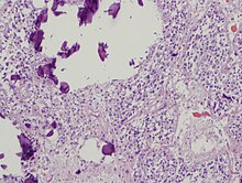
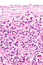
Tumors[]
Tumors of the pineal gland are called pinealomas. These tumors are rare and 50% to 70% are germinomas that arise from sequestered embryonic germ cells. Histologically they are similar to testicular seminomas and ovarian dysgerminomas.[38]
A pineal tumor can compress the superior colliculi and pretectal area of the dorsal midbrain, producing Parinaud's syndrome. Pineal tumors also can cause compression of the cerebral aqueduct, resulting in a noncommunicating hydrocephalus. Other manifestations are the consequence of their pressure effects and consist of visual disturbances, headache, mental deterioration, and sometimes dementia-like behaviour.[39]
These neoplasms are divided into three categories: pineoblastomas, pineocytomas, and mixed tumors, based on their level of differentiation, which, in turn, correlates with their neoplastic aggressiveness.[40] The clinical course of patients with pineocytomas is prolonged, averaging up to several years.[41] The position of these tumors makes them difficult to remove surgically.
Other conditions[]
The morphology of the pineal gland differs markedly in different pathological conditions. For instance, it is known that its volume is reduced both in obese patients as well as patients with primary insomnia.[42]
Other animals[]
Most living vertebrates have pineal glands. It is likely that the common ancestor of all vertebrates had a pair of photosensory organs on the top of its head, similar to the arrangement in modern lampreys.[43] Some extinct Devonian fishes have two parietal foramina in their skulls,[44][45] suggesting an ancestral bilaterality of parietal eyes. The parietal eye and the pineal gland of living tetrapods are probably the descendants of the left and right parts of this organ, respectively.[46]
During embryonic development, the parietal eye and the pineal organ of modern lizards[47] and tuataras[48] form together from a pocket formed in the brain ectoderm. The loss of parietal eyes in many living tetrapods is supported by developmental formation of a paired structure that subsequently fuses into a single pineal gland in developing embryos of turtles, snakes, birds, and mammals.[49]
The pineal organs of mammals fall into one of three categories based on shape. Rodents have more structurally complex pineal glands than other mammals.[50]
Crocodilians and some tropical lineages of mammals (some xenarthrans (sloths), pangolins, sirenians (manatees & dugongs), and some marsupials (sugar gliders) have lost both their parietal eye and their pineal organ.[51][52][50] Polar mammals, such as walruses and some seals, possess unusually large pineal glands.[51]
All amphibians have a pineal organ, but some frogs and toads also have what is called a "frontal organ", which is essentially a parietal eye.[53]
Pinealocytes in many non-mammalian vertebrates have a strong resemblance to the photoreceptor cells of the eye. Evidence from morphology and developmental biology suggests that pineal cells possess a common evolutionary ancestor with retinal cells.[54]
Pineal cytostructure seems to have evolutionary similarities to the retinal cells of the lateral eyes.[54] Modern birds and reptiles express the phototransducing pigment melanopsin in the pineal gland. Avian pineal glands are thought to act like the suprachiasmatic nucleus in mammals.[55] The structure of the pineal eye in modern lizards and tuatara is analogous to the cornea, lens, and retina of the lateral eyes of vertebrates.[49]
In most vertebrates, exposure to light sets off a chain reaction of enzymatic events within the pineal gland that regulates circadian rhythms.[56] In humans and other mammals, the light signals necessary to set circadian rhythms are sent from the eye through the retinohypothalamic system to the suprachiasmatic nuclei (SCN) and the pineal gland.
The fossilized skulls of many extinct vertebrates have a pineal foramen (opening), which in some cases is larger than that of any living vertebrate.[57] Although fossils seldom preserve deep-brain soft anatomy, the brain of the Russian fossil bird Cerebavis cenomanica from Melovatka, about 90 million years old, shows a relatively large parietal eye and pineal gland.[58]
Rick Strassman, an author and Clinical Associate Professor of Psychiatry at the University of New Mexico School of Medicine, has theorised that the human pineal gland is capable of producing the hallucinogen N,N-Dimethyltryptamine (DMT) under certain circumstances.[59] In 2013 he and other researchers first reported DMT in the pineal gland microdialysate of rodents.[60]
Society and culture[]
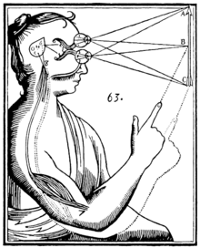
Seventeenth-century philosopher and scientist René Descartes was highly interested in anatomy and physiology. He discussed the pineal gland both in his first book, the Treatise of Man (written before 1637, but only published posthumously 1662/1664), and in his last book, The Passions of the Soul (1649) and he regarded it as "the principal seat of the soul and the place in which all our thoughts are formed."[61] In the Treatise of Man, Descartes described conceptual models of man, namely creatures created by God, which consist of two ingredients, a body and a soul.[61][62] In the Passions, Descartes split man up into a body and a soul and emphasized that the soul is joined to the whole body by "a certain very small gland situated in the middle of the brain's substance and suspended above the passage through which the spirits in the brain's anterior cavities communicate with those in its posterior cavities". Descartes attached significance to the gland because he believed it to be the only section of the brain to exist as a single part rather than one-half of a pair. Some of Descartes's basic anatomical and physiological assumptions were totally mistaken, not only by modern standards, but also in light of what was already known in his time.[61]
The notion of a "pineal-eye" is central to the philosophy of the French writer Georges Bataille, which is analyzed at length by literary scholar Denis Hollier in his study Against Architecture. In this work Hollier discusses how Bataille uses the concept of a "pineal-eye" as a reference to a blind-spot in Western rationality, and an organ of excess and delirium.[63] This conceptual device is explicit in his surrealist texts, The Jesuve and The Pineal Eye.[64]
In the late 19th century Madame Blavatsky (who founded theosophy) identified the pineal gland with the Hindu concept of the third eye, or the Ajna chakra. This association is still popular today.[61]
In the short story "From Beyond" by H. P. Lovecraft, a scientist creates an electronic device that emits a resonance wave, which stimulates an affected person's pineal gland, thereby allowing them to perceive planes of existence outside the scope of accepted reality, a translucent, alien environment that overlaps our own recognized reality. It was adapted as a film of the same name in 1986. The 2013 horror film Banshee Chapter is heavily influenced by this short story.
History[]
The secretory activity of the pineal gland is only partially understood. Its location deep in the brain suggested to philosophers throughout history that it possesses particular importance. This combination led to its being regarded as a "mystery" gland with mystical, metaphysical, and occult theories surrounding its perceived functions.
The pineal gland was originally believed to be a "vestigial remnant" of a larger organ. In 1917, it was known that extract of cow pineals lightened frog skin. Dermatology professor Aaron B. Lerner and colleagues at Yale University, hoping that a substance from the pineal might be useful in treating skin diseases, isolated and named the hormone melatonin in 1958.[65] The substance did not prove to be helpful as intended, but its discovery helped solve several mysteries such as why removing the rat's pineal accelerated ovary growth, why keeping rats in constant light decreased the weight of their pineals, and why pinealectomy and constant light affect ovary growth to an equal extent; this knowledge gave a boost to the then new field of chronobiology.[66]
Additional images[]
The pineal body is labeled in these images.

Mesal aspect of a brain sectioned in the median sagittal plane.

Dissection showing the ventricles of the brain.

Hind- and mid-brains; antero-lateral view.
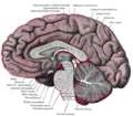
Median sagittal section of brain.

Pineal gland

Brainstem. Posterior view.


See also[]
References[]
- ^ Jump up to: a b c "Pineal (as an adjective)". Online Etymology Dictionary, Douglas Harper. 2018. Retrieved 27 October 2018.
- ^ Macchi MM, Bruce JN (2004). "Human pineal physiology and functional significance of melatonin". Frontiers in Neuroendocrinology. 25 (3–4): 177–95. doi:10.1016/j.yfrne.2004.08.001. PMID 15589268. S2CID 26142713.
- ^ Arendt J, Skene DJ (February 2005). "Melatonin as a chronobiotic". Sleep Medicine Reviews. 9 (1): 25–39. doi:10.1016/j.smrv.2004.05.002. PMID 15649736.
Exogenous melatonin has acute sleepiness-inducing and temperature-lowering effects during 'biological daytime', and when suitably timed (it is most effective around dusk and dawn) it will shift the phase of the human circadian clock (sleep, endogenous melatonin, cortisol) to earlier (advance phase shift) or later (delay phase shift) times.
- ^ Gross PM, Weindl A (December 1987). "Peering through the windows of the brain". Journal of Cerebral Blood Flow and Metabolism. 7 (6): 663–72. doi:10.1038/jcbfm.1987.120. PMID 2891718. S2CID 18748366.
- ^ Ooka-Souda S, Kadota T, Kabasawa H (December 1993). "The preoptic nucleus: the probable location of the circadian pacemaker of the hagfish, Eptatretus burgeri". Neuroscience Letters. 164 (1–2): 33–6. doi:10.1016/0304-3940(93)90850-K. PMID 8152610. S2CID 40006945.
- ^ Jump up to: a b Vernadakis AJ, Bemis WE, Bittman EL (April 1998). "Localization and partial characterization of melatonin receptors in amphioxus, hagfish, lamprey, and skate". General and Comparative Endocrinology. 110 (1): 67–78. doi:10.1006/gcen.1997.7042. PMID 9514841.
- ^ Erlich SS, Apuzzo ML (September 1985). "The pineal gland: anatomy, physiology, and clinical significance". Journal of Neurosurgery. 63 (3): 321–41. doi:10.3171/jns.1985.63.3.0321. PMID 2862230. S2CID 29929205.
- ^ Eakin, Richard M. (1973). The Third Eye. Berkeley: University of California Press.
- ^ Lokhorst, Gert-Jan (2018), "Descartes and the Pineal Gland", in Zalta, Edward N. (ed.), The Stanford Encyclopedia of Philosophy (Winter 2018 ed.), Metaphysics Research Lab, Stanford University, retrieved 17 December 2019
- ^ Bowen R. "The Pineal Gland and Melatonin". Archived from the original on 24 November 2011. Retrieved 14 October 2011.
- ^ Chen CY, Chen FH, Lee CC, Lee KW, Hsiao HS (October 1998). "Sonographic characteristics of the cavum velum interpositum" (PDF). AJNR. American Journal of Neuroradiology. 19 (9): 1631–5. PMC 8337493. PMID 9802483.
- ^ Dorland's. Illustrated Medical Dictionary. Elsevier Saunders. p. 1607. ISBN 978-1-4160-6257-8.
- ^ Pritchard TC, Alloway KD (1999). Medical Neuroscience (Google books preview). Hayes Barton Press. pp. 76–77. ISBN 978-1-889325-29-3. Retrieved 8 February 2009.
- ^ Arendt J: Melatonin and the Mammalian Pineal Gland, ed 1. London. Chapman & Hall, 1995, p 17
- ^ Møller M, Baeres FM (July 2002). "The anatomy and innervation of the mammalian pineal gland". Cell and Tissue Research. 309 (1): 139–50. doi:10.1007/s00441-002-0580-5. PMID 12111544. S2CID 25719864.
- ^ Kleinschmidt-DeMasters BK, Prayson RA (November 2006). "An algorithmic approach to the brain biopsy--part I". Archives of Pathology & Laboratory Medicine. 130 (11): 1630–8. doi:10.5858/2006-130-1630-AAATTB. PMID 17076524.
- ^ Schmidt F, Penka B, Trauner M, Reinsperger L, Ranner G, Ebner F, Waldhauser F (April 1995). "Lack of pineal growth during childhood". The Journal of Clinical Endocrinology and Metabolism. 80 (4): 1221–5. doi:10.1210/jcem.80.4.7536203. PMID 7536203.
- ^ Sumida M, Barkovich AJ, Newton TH (February 1996). "Development of the pineal gland: measurement with MR". AJNR. American Journal of Neuroradiology. 17 (2): 233–6. PMC 8338352. PMID 8938291.
- ^ Tapp E, Huxley M (September 1971). "The weight and degree of calcification of the pineal gland". The Journal of Pathology. 105 (1): 31–9. doi:10.1002/path.1711050105. PMID 4943068. S2CID 38346296.
- ^ Tapp E, Huxley M (October 1972). "The histological appearance of the human pineal gland from puberty to old age". The Journal of Pathology. 108 (2): 137–44. doi:10.1002/path.1711080207. PMID 4647506. S2CID 28529644.
- ^ "Melatonin Production and Age". Chronobiology. Medichron Publications.
- ^ Snelson CD, Santhakumar K, Halpern ME, Gamse JT (May 2008). "Tbx2b is required for the development of the parapineal organ". Development. 135 (9): 1693–702. doi:10.1242/dev.016576. PMC 2810831. PMID 18385257.
- ^ Snelson CD, Burkart JT, Gamse JT (December 2008). "Formation of the asymmetric pineal complex in zebrafish requires two independently acting transcription factors". Developmental Dynamics. 237 (12): 3538–44. doi:10.1002/dvdy.21607. PMC 2810829. PMID 18629869.
- ^ Snelson CD, Gamse JT (June 2009). "Building an asymmetric brain: development of the zebrafish epithalamus". Seminars in Cell & Developmental Biology. 20 (4): 491–7. doi:10.1016/j.semcdb.2008.11.008. PMC 2729063. PMID 19084075.
- ^ Axelrod J (September 1970). "The pineal gland". Endeavour. 29 (108): 144–8. PMID 4195878.
- ^ Lowrey PL, Takahashi JS (2000). "Genetics of the mammalian circadian system: Photic entrainment, circadian pacemaker mechanisms, and posttranslational regulation". Annual Review of Genetics. 34 (1): 533–562. doi:10.1146/annurev.genet.34.1.533. PMID 11092838.
- ^ Callaway JC, Gyntber J, Poso A, Airaksinen MM, Vepsäläinen J (1994). "The pictet-spengler reaction and biogenic tryptamines: Formation of tetrahydro-β-carbolines at physiologicalpH". Journal of Heterocyclic Chemistry. 31 (2): 431–435. doi:10.1002/jhet.5570310231.
- ^ Jump up to: a b Motta M, Fraschini F, Martini L (November 1967). "Endocrine effects of pineal gland and of melatonin". Proceedings of the Society for Experimental Biology and Medicine. 126 (2): 431–5. doi:10.3181/00379727-126-32468. PMID 6079917. S2CID 28964258.
- ^ Naidoo V, Naidoo S, Mahabeer R, Raidoo DM (May 2004). "Cellular distribution of the endothelin system in the human brain". Journal of Chemical Neuroanatomy. 27 (2): 87–98. doi:10.1016/j.jchemneu.2003.12.002. PMID 15121213. S2CID 39053816.
- ^ Gross PM, Wainman DS, Chew BH, Espinosa FJ, Weaver DF (March 1993). "Calcium-mediated metabolic stimulation of neuroendocrine structures by intraventricular endothelin-1 in conscious rats". Brain Research. 606 (1): 135–42. doi:10.1016/0006-8993(93)91581-C. PMID 8461995. S2CID 12713010.
- ^ Sharan K, Lewis K, Furukawa T, Yadav VK (September 2017). "Regulation of bone mass through pineal-derived melatonin-MT2 receptor pathway". Journal of Pineal Research. 63 (2): e12423. doi:10.1111/jpi.12423. PMC 5575491. PMID 28512916.
- ^ Jump up to: a b c Zimmerman RA (1982). "Age-Related Incidence of Pineal Calcification Detected by Computed Tomography" (PDF). Radiology. Radiological Society of North America. 142 (3): 659–62. doi:10.1148/radiology.142.3.7063680. PMID 7063680. Archived from the original (PDF) on 24 March 2012. Retrieved 21 June 2012.
- ^ "The Pineal Body". Human Anatomy (Gray's Anatomy). Archived from the original on 26 August 2011. Retrieved 7 September 2011.
- ^ Kunz, D.; Schmitz, S.; Mahlberg, R.; Mohr, A.; Stöter, C.; Wolf, K. J.; Herrmann, W. M. (21 December 1999). "A new concept for melatonin deficit: on pineal calcification and melatonin excretion". Neuropsychopharmacology. 21 (6): 765–772. doi:10.1016/S0893-133X(99)00069-X. ISSN 0893-133X. PMID 10633482. S2CID 20184373.
- ^ Tan, Dun Xian; Xu, Bing; Zhou, Xinjia; Reiter, Russel J. (31 January 2018). "Pineal Calcification, Melatonin Production, Aging, Associated Health Consequences and Rejuvenation of the Pineal Gland". Molecules. 23 (2): 301. doi:10.3390/molecules23020301. ISSN 1420-3049. PMC 6017004. PMID 29385085.
- ^ Del Brutto, Oscar H.; Mera, Robertino M.; Lama, Julio; Zambrano, Mauricio; Castillo, Pablo R. (November 2014). "Pineal gland calcification is not associated with sleep-related symptoms. A population-based study in community-dwelling elders living in Atahualpa (rural coastal Ecuador)". Sleep Medicine. 15 (11): 1426–1427. doi:10.1016/j.sleep.2014.07.008. ISSN 1878-5506. PMID 25277665.
- ^ Mahlberg R, Kienast T, Hädel S, Heidenreich JO, Schmitz S, Kunz D (April 2009). "Degree of pineal calcification (DOC) is associated with polysomnographic sleep measures in primary insomnia patients". Sleep Medicine. 10 (4): 439–445. doi:10.1016/j.sleep.2008.05.003. PMID 18755628.
- ^ Kumar V, Abbas AK, Aster JC (5 September 2014). Robbins & Cotran Pathologic Basis of Disease. p. 1137. ISBN 9780323296359.
- ^ Bruce J. "Pineal Tumours". eMedicine. Archived from the original on 5 September 2015. Retrieved 25 September 2015.
- ^ "Pineal Tumours". American Brain Tumour Association. Archived from the original on 26 September 2015. Retrieved 25 September 2015.
- ^ Clark AJ, Sughrue ME, Ivan ME, Aranda D, Rutkowski MJ, Kane AJ, Chang S, Parsa AT (November 2010). "Factors influencing overall survival rates for patients with pineocytoma". Journal of Neuro-Oncology. 100 (2): 255–60. doi:10.1007/s11060-010-0189-6. PMC 2995321. PMID 20461445.
- ^ Tan, Dun Xian; Xu, Bing; Zhou, Xinjia; Reiter, Russel J. (February 2018). "Pineal Calcification, Melatonin Production, Aging, Associated Health Consequences and Rejuvenation of the Pineal Gland". Molecules. 23 (2): 301. doi:10.3390/molecules23020301. PMC 6017004. PMID 29385085. S2CID 25611663.
- ^ Cole WC, Youson JH (October 1982). "Morphology of the pineal complex of the anadromous sea lamprey, Petromyzon marinus L". The American Journal of Anatomy. 165 (2): 131–63. doi:10.1002/aja.1001650205. PMID 7148728.
- ^ Cope ED (1888). "The pineal eye in extinct vertebrates". American Naturalist. 22 (262): 914–917. doi:10.1086/274797. S2CID 85280810.
- ^ Schultze H (1993). "Patterns of diversity in the skulls of jawed fishes". In Hanken J, Hall BK (eds.). The Skull. 2: Patterns of Structural and Systematic Diversity. Chicago, Illinois: University of Chicago Press. pp. 189–254. ISBN 9780226315683. Retrieved 2 February 2017.
- ^ Dodt E (1973). "The parietal eye (pineal and parietal organs) of lower vertebrates". Visual Centers in the Brain. Springer. pp. 113–140.
- ^ Tosini G (1997). "The pineal complex of reptiles: physiological and behavioral roles". Ethology Ecology & Evolution. 9 (4): 313–333. doi:10.1080/08927014.1997.9522875.
- ^ Dendy A (1911). "On the structure, development and morphological interpretation of the pineal organs and adjacent parts of the brain in the tuatara (Sphenodon punctatus)". Philosophical Transactions of the Royal Society of London B. 201 (274–281): 227–331. doi:10.1098/rstb.1911.0006.
- ^ Jump up to: a b Quay WB (1979). "The parietal eye–pineal complex". In Gans C, Northcutt RG, Ulinski P (eds.). Biology of the Reptilia. Volume 9. Neurology A. London: Academic Press. pp. 245–406. Archived from the original on 3 February 2017.
- ^ Jump up to: a b Vollrath L (1979). Comparative morphology of the vertebrate pineal complex. Progress in Brain Research. 52. pp. 25–38. doi:10.1016/S0079-6123(08)62909-X. ISBN 9780444801142. PMID 398532.
- ^ Jump up to: a b Ralph CL (December 1975). "The pineal gland and geographical distribution of animals". International Journal of Biometeorology. 19 (4): 289–303. Bibcode:1975IJBm...19..289R. doi:10.1007/bf01451040. PMID 1232070. S2CID 30406445.
- ^ Ralph C, Young S, Gettinger R, O'Shea TJ (1985). "Does the manatee have a pineal body?". Acta Zoologica. 66: 55–60. doi:10.1111/j.1463-6395.1985.tb00647.x.
- ^ Adler K (1976). "Extraocular photoreception in amphibians". Photochemistry and Photobiology. 23 (4): 275–298. doi:10.1111/j.1751-1097.1976.tb07250.x. PMID 775500. S2CID 33692776.
- ^ Natesan A, Geetha L, Zatz M (July 2002). "Rhythm and soul in the avian pineal". Cell and Tissue Research. 309 (1): 35–45. doi:10.1007/s00441-002-0571-6. PMID 12111535. S2CID 26023207.
- ^ Moore RY, Heller A, Wurtman RJ, Axelrod J (January 1967). "Visual pathway mediating pineal response to environmental light". Science. 155 (3759): 220–3. Bibcode:1967Sci...155..220M. doi:10.1126/science.155.3759.220. PMID 6015532. S2CID 44377291.
- ^ Edinger T (1955). "The size of parietal foramen and organ in reptiles: a rectification". Bulletin of the Museum of Comparative Zoology. 114: 1–34. Archived from the original on 1 December 2017.
- ^ Kurochkin EN, Dyke GJ, Saveliev SV, Pervushov EM, Popov EV (June 2007). "A fossil brain from the Cretaceous of European Russia and avian sensory evolution". Biology Letters. 3 (3): 309–13. doi:10.1098/rsbl.2006.0617. PMC 2390680. PMID 17426009.
- ^ Strassman R (2000). DMT: The Spirit Molecule. Inner Traditions. ISBN 978-1594779732. Archived from the original on 6 January 2017.
- ^ Barker SA, Borjigin J, Lomnicka I, Strassman R (December 2013). "LC/MS/MS analysis of the endogenous dimethyltryptamine hallucinogens, their precursors, and major metabolites in rat pineal gland microdialysate" (PDF). Biomedical Chromatography. 27 (12): 1690–700. doi:10.1002/bmc.2981. hdl:2027.42/101767. PMID 23881860.
- ^ Jump up to: a b c d (2015). Descartes and the Pineal Gland. Stanford: The Stanford Encyclopedia of Philosophy.
- ^ Descartes R. "The Passions of the Soul" excerpted from "Philosophy of the Mind," Chalmers, D. New York: Oxford University Press, Inc.; 2002. ISBN 978-0-19-514581-6
- ^ Hollier, D, Against Architecture: The Writings of Georges Bataille, trans. Betsy Wing, MIT, 1989.
- ^ Bataille, G, Visions of Excess: Selected Writings, 1927–1939 (Theory and History of Literature, Vol 14), trans. Allan Stoekl et al., Manchester University Press, 1985
- ^ Lerner AB, Case JD, Takahashi Y (July 1960). "Isolation of melatonin and 5-methoxyindole-3-acetic acid from bovine pineal glands". The Journal of Biological Chemistry. 235 (7): 1992–7. doi:10.1016/S0021-9258(18)69351-2. PMID 14415935.
- ^ Coates PM, Blackman MR, Cragg GM, Levine M, Moss J, White JD (2005). Encyclopedia of Dietary Supplements. CRC Press. p. 457. ISBN 978-0-8247-5504-1. Retrieved 31 March 2009.
External links[]
| Wikimedia Commons has media related to Pineal gland. |
| Look up pineal gland in Wiktionary, the free dictionary. |
| show Authority control |
|---|
- Circadian rhythm
- Endocrine system
- Epithalamus
- Glands
- Human head and neck
- Sleep physiology