External vertebral venous plexuses
| External vertebral venous plexuses | |
|---|---|
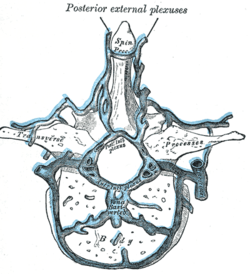 Transverse section of a thoracic vertebra, showing the vertebral venous plexuses. | |
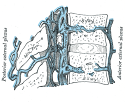 Median sagittal section of two thoracic vertebrae, showing the vertebral venous plexuses. | |
| Details | |
| Identifiers | |
| Latin | plexus venosi vertebrales externi |
| TA98 | A12.3.07.019 A12.3.07.020 |
| TA2 | 4951, 4952 |
| FMA | 12851 |
| Anatomical terminology | |
The external vertebral venous plexuses (extraspinal veins) best marked in the cervical region, consist of anterior and posterior plexuses which anastomose freely with each other.
- The anterior external plexuses lie in front of the bodies of the vertebræ, communicate with the basivertebral and intervertebral veins, and receive tributaries from the vertebral bodies.
- The posterior external plexuses are placed partly on the posterior surfaces of the vertebral arches and their processes, and partly between the deep dorsal muscles.
They are best developed in the cervical region, and there anastomose with the vertebral, occipital, and deep cervical veins.
References[]
![]() This article incorporates text in the public domain from page 668 of the 20th edition of Gray's Anatomy (1918)
This article incorporates text in the public domain from page 668 of the 20th edition of Gray's Anatomy (1918)
External links[]
- Atlas image: abdo_wall77 at the University of Michigan Health System - "Venous Drainage of the Vertebral Column"
| Authority control: Scientific databases |
|---|
This cardiovascular system article is a stub. You can help Wikipedia by . |
Categories:
- Wikipedia articles incorporating text from the 20th edition of Gray's Anatomy (1918)
- Veins of the torso
- Cardiovascular system stubs