Brachiocephalic vein
| Brachiocephalic vein | |
|---|---|
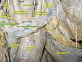
 The thyroid gland and its relations. (Label for "Right innom. vein" and "Left innom. vein" visible at bottom center.) | |
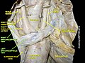
 The arch of the aorta, and its branches. (Right innom. vein labeled at upper right; left innominate vein labeled at center top.) | |
| Details | |
| Source | Internal jugular subclavian superior intercostal vertebral inferior thyroid |
| Drains to | Superior vena cava |
| Artery | Brachiocephalic artery |
| Identifiers | |
| Latin | vena brachiocephalica vena anonyma |
| MeSH | D016121 |
| TA98 | A12.3.04.001 |
| TA2 | 4772 |
| FMA | 4723 |
| Anatomical terminology | |
The left and right brachiocephalic veins (or innominate veins) are major veins in the upper chest, formed by the union of each corresponding internal jugular vein and subclavian vein.[1] This is at the level of the sternoclavicular joint.[2] The left brachiocephalic vein is nearly always longer than the right.[3]
These veins merge to form the superior vena cava, a great vessel, posterior to the junction of the first costal cartilage with the manubrium of the sternum.[4][5]
The brachiocephalic veins are the major veins returning blood to the superior vena cava.[3]
Tributaries[]
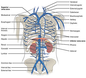
The brachiocephalic vein is formed by the confluence of the subclavian and internal jugular veins.[1] In addition it receives drainage from:
- Left and right internal thoracic vein (Also called internal mammary veins): drain into the inferior border of their corresponding vein
- Left and right inferior thyroid veins: drain into the superior aspect of their corresponding veins near the confluence
- Left and right vertebral vein
- Left superior intercostal vein: drains into the left brachiocephalic vein[6]
Embryological origin[]
The left brachiocephalic vein forms from the anastomosis formed between the left and right anterior cardinal veins when the caudal portion of the left anterior cardinal vein degenerates.
Additional images[]
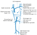
Diagram showing completion of development of the parietal veins.
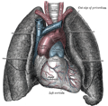
Front view of heart and lungs.
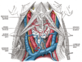
The fascia and middle thyroid veins.

Right Brachiocephalic vein
Right& Left Brachiocephalic vein
Right& Left Brachiocephalic vein

The brachiocephalic veins, superior vena cava, inferior vena cava, azygos vein and their tributaries.


References[]
- ^ a b Gupta, Arjun; Kim, D. Nathan; Kalva, Sanjeeva; Reznik, Scott; Johnson, David H. (2020-01-01), Niederhuber, John E.; Armitage, James O.; Kastan, Michael B.; Doroshow, James H. (eds.), "53 - Superior Vena Cava Syndrome", Abeloff's Clinical Oncology (Sixth Edition), Philadelphia: Elsevier, pp. 775–785.e2, doi:10.1016/b978-0-323-47674-4.00053-0, ISBN 978-0-323-47674-4, retrieved 2020-11-20
- ^ Chitnis, Cumberbatch, Gankande. Practice Papers for MCEM Part A, Wiley-Blackwell 2010[page needed]
- ^ a b Fligner, Corinne L.; Clark, John I.; Clark, Judy M.; Larson, Lyle W.; Poole, Jeanne E. (2018-01-01), Poole, Jeanne E.; Larson, Lyle W. (eds.), "2 - Surgical Anatomy for the Implanting Physician", Surgical Implantation of Cardiac Rhythm Devices, Elsevier, pp. 13–58, doi:10.1016/b978-0-323-40126-5.00002-1, ISBN 978-0-323-40126-5, retrieved 2020-11-20
- ^ Mozes, GEZA; Gloviczki, PETER (2007-01-01), Bergan, John J. (ed.), "CHAPTER 2 - Venous Embryology and Anatomy", The Vein Book, Burlington: Academic Press, pp. 15–25, doi:10.1016/b978-012369515-4/50005-3, ISBN 978-0-12-369515-4, retrieved 2020-11-20
- ^ Long, Chandler A.; Kwolek, Christopher J.; Watkins, Michael T. (2013-01-01), Creager, Mark A.; Beckman, Joshua A.; Loscalzo, Joseph (eds.), "Chapter 61 - Vascular Trauma", Vascular Medicine: A Companion to Braunwald's Heart Disease (Second Edition), Philadelphia: W.B. Saunders, pp. 739–754, doi:10.1016/b978-1-4377-2930-6.00061-6, ISBN 978-1-4377-2930-6, retrieved 2020-11-20
- ^ Ryan, McNicholas & Eustace "Anatomy for Diagnostic Imaging: 3rd Edition"[page needed]
| Authority control: Scientific databases |
|---|
This cardiovascular system article is a stub. You can help Wikipedia by . |
- Veins of the torso
- Cardiovascular system stubs