HeLa


HeLa (/ˈhiːlɑː/; also Hela or hela) is an immortal cell line used in scientific research. It is the oldest and most commonly used human cell line.[1] The line is named after and derived from cervical cancer cells taken on February 8, 1951,[2] from Henrietta Lacks, a 31-year-old African-American mother of five, who died of cancer on October 4, 1951.[3] The cell line was found to be remarkably durable and prolific, which allows it to be used extensively in scientific study.[4][5]
The cells from Lacks's cancerous cervical tumor were taken without her knowledge or consent, which was common practice at the time.[6] Cell biologist George Otto Gey found that they could be kept alive,[7] and developed a cell line. Previously, cells cultured from other human cells would only survive for a few days. Cells from Lacks's tumor behaved differently.
History[]
Origin[]
In 1951, a patient named Henrietta Lacks was admitted to the Johns Hopkins Hospital with symptoms of irregular vaginal bleeding, and was subsequently treated for cervical cancer.[8] Her cervical biopsy supplied samples of tissue for clinical evaluation and research by Dr. George Otto Gey, head of the Tissue Culture Laboratory, as was done with other surgical procedures. Gey's lab assistant Mary Kubicek used the roller-tube technique to place the cells into culture.[8] It was observed that the cells grew robustly, doubling every 20–24 hours unlike previous specimens that died out.[9]
The cells were propagated by Gey shortly before Lacks died of her cancer in 1951. This was the first human cell line to prove successful in vitro, which was a scientific achievement with profound future benefit to medical research. Gey freely donated these cells along with the tools and processes that his lab developed to any scientist requesting them simply for the benefit of science. Neither Lacks nor her family gave permission to harvest the cells but, at that time, permission was neither required nor customarily sought.[10] The cells were later commercialized, although never patented in their original form. There was no requirement at that time to inform patients or their relatives about such matters because discarded material or material obtained during surgery, diagnosis, or therapy was the property of the physician or the medical institution.
As was custom for Gey's lab assistant, the culture was named after the first two letters of the Henrietta Lacks' first and last name.[8] Before a leak to the public in the 1970s which revealed her true name, the "HeLa" cell line was mistakenly believed to have been named after a "Helen Lane" or "Helen Larson".[5][11]
Starting in the 1970s, the Lacks family was contacted by researchers trying to find out why the HeLa cells had contaminated other cell lines in laboratories.[12] These cells are treated as cancer cells, as they are descended from a biopsy taken from a visible lesion on the cervix as part of Lacks's diagnosis of cancer.[13]
HeLa cells, like other cell lines, are termed "immortal" in that they can divide an unlimited number of times in a laboratory cell culture plate as long as fundamental cell survival conditions are met (i.e. being maintained and sustained in a suitable environment). There are many strains of HeLa cells as they continue to mutate in cell cultures, but all HeLa cells are descended from the same tumor cells removed from Lacks. The total number of HeLa cells that have been propagated in cell culture far exceeds the total number of cells that were in Henrietta Lacks's body.[14]
Controversy[]
Lacks's case is one of many examples of the lack of informed consent in 20th century medicine. Communication between tissue donors and doctors was virtually nonexistent (i.e. cells were taken without patient consent, nor were they told what the cells would be used for). Lacks's family also had no access to her patient files and had no say in who received HeLa cells or what they would be used for. Additionally, as HeLa cells were popularized and used more frequently throughout the scientific community, Lacks's relatives received no financial benefit and continued to live with limited access to healthcare.[15]
This issue of who owns tissue samples taken for research was brought up in the Supreme Court of California case of Moore v. Regents of the University of California. The court ruled that a person's discarded tissue and cells are not his or her property and can be commercialized.[16]
Lacks's case influenced the establishment of the Common Rule in 1981. The Common Rule enforces informed consent by ensuring that doctors inform patients if they plan to use any details of the patient's case in research and give them the choice of disclosing the details or not. Tissues connected to their donors' names are also strictly regulated under this rule, and samples are no longer named using donor initials, but rather by code numbers.[16] To further resolve the issue of patient privacy, Johns Hopkins established a joint committee with the NIH and several of Lacks's family members to determine who receives access to Henrietta Lacks's genome.[17]
Use in research[]
HeLa cells were the first human cells to be successfully cloned in 1953 by Theodore Puck and Philip I. Marcus at the University of Colorado, Denver.[18] Since that time, HeLa cells have "continually been used for research into cancer, AIDS, the effects of radiation and toxic substances, gene mapping, and countless other scientific pursuits."[19] According to author Rebecca Skloot, by 2009, "more than 60,000 scientific articles had been published about research done on HeLa, and that number was increasing steadily at a rate of more than 300 papers each month."[16]
Polio eradication[]
HeLa cells were used by Jonas Salk to test the first polio vaccine in the 1950s. They were observed to be easily infected by poliomyelitis, causing infected cells to die.[20] This made HeLa cells highly desirable for polio vaccine testing since results could be easily obtained. A large volume of HeLa cells were needed for the testing of Salk's polio vaccine, prompting the National Foundation for Infantile Paralysis (NFIP) to find a facility capable of mass-producing HeLa cells.[21] In the spring of 1953, a cell culture factory was established at Tuskegee University to supply Salk and other labs with HeLa cells.[22] Less than a year later, Salk's vaccine was ready for human trials.[23]
Virology[]
HeLa cells have been used in testing how parvovirus infects cells of humans, dogs, and cats.[24] These cells have also been used to study viruses such as the oropouche virus (OROV). OROV causes the disruption of cells in culture, where cells begin to degenerate shortly after they are infected, causing viral induction of apoptosis.[25] HeLa cells have been used to study the expression of the papillomavirus E2 and apoptosis.[26] HeLa cells have also been used to study canine distemper virus' ability to induce apoptosis in cancer cell lines,[27] which could play an important role in developing treatments for tumor cells resistant to radiation and chemotherapy.[27]
HeLa cells have also been instrumental in the development of human papilloma virus (HPV) vaccines. In the 1980s, Harald zur Hausen found that Lacks's cells from the original biopsy contained HPV-18, which was later found to be the cause of the aggressive cancer that killed Henrietta Lacks. His work in linking HPV with cervical cancer won him a Nobel Prize and led to the development of HPV vaccines that are predicted to reduce the number of deaths from cervical cancer by 70%.[28]
Over the years, HeLa cells have been infected with various types of viruses including HIV, Zika, herpes, and mumps to test and develop new vaccines and drugs. Dr. Richard Axel discovered that by adding the CD4 protein to HeLa cells, they were able to be infected with HIV, allowing the virus to be studied.[29] In 1979, scientists learned that the measles virus constantly mutates when it infects HeLa cells[30] and in 2019, found that Zika cannot multiply in HeLa cells.[31]
Cancer[]
HeLa cells have been used in a number of cancer studies, including those involving sex steroid hormones such as estradiol, estrogen, and estrogen receptors, along with estrogen-like compounds such as quercetin and its cancer reducing properties.[32] There have also been studies on HeLa cells, the effects of flavonoids and antioxidants with estradiol on cancer cell proliferation.
In 2011, HeLa cells were used in tests of novel heptamethine dyes IR-808 and other analogs which are currently being explored for their unique uses in medical diagnostics, the development of theranostics, the individualized treatment of cancer patients with the aid of PDT, co-administration with other drugs, and irradiation.[33][34] HeLa cells have been used in research involving fullerenes to induce apoptosis as a part of photodynamic therapy, as well as in in vitro cancer research using cell lines.[35] Further HeLa cells have also been used to define cancer markers in RNA, and have been used to establish an RNAi Based Identification System and Interference of Specific Cancer Cells.[36]
HeLa was shown in 2014 to be viable cell line for tumor xenografts in C57BL/6 nude mice,[37] and was subsequently used to examine the in vivo effects of fluoxetine and cisplatin on cervical cancer.
Genetics[]
In 1953, a lab mistake involving mixing HeLa cells with the wrong liquid allowed researchers to see and count each chromosome clearly in the HeLa cells they were working with for the first time. The accidental discovery led scientists Joe Hin Tjio and Albert Levan to develop better techniques for staining and counting chromosomes.[28] They were the first to accurately describe that humans have 23 pairs of chromosomes rather than 24, as was previously believed. This was important for the study of developmental disorders such as Down syndrome that involved the number of chromosomes.
In 1965, Henry Harris and John Watkins created the first human-animal hybrid by fusing HeLa cells with mouse embryo cells. This enabled advancements in mapping genes to specific chromosomes, which would eventually lead to the Human Genome Project.[28]
Space microbiology[]
In the 1960s, HeLa cells were sent on the Soviet satellite "Sputnik-6" and human space missions to determine the long term effects of space travel on living cells and tissue. Scientists discovered that HeLa cells divided even more quickly in zero gravity.[38]
Analysis[]
Telomerase[]
The HeLa cell line was derived for use in cancer research. These cells proliferate abnormally rapidly, even compared to other cancer cells. Like many other cancer cells,[39] HeLa cells have an active version of telomerase during cell division,[40] which copies telomeres over and over again. This prevents the incremental shortening of telomeres that is implicated in aging and eventual cell death. In this way, the cells circumvent the Hayflick limit, which is the limited number of cell divisions that most normal cells can undergo before becoming senescent. The result is unlimited cell division and immortality.
Chromosome number[]
Horizontal gene transfer from human papillomavirus 18 (HPV18) to human cervical cells created the HeLa genome, which is different from Henrietta Lacks' genome in various ways, including its number of chromosomes. HeLa cells are rapidly dividing cancer cells, and the number of chromosomes varied during cancer formation and cell culture. The current estimate (excluding very tiny fragments) is a "hypertriploid chromosome number (3n+)" which means 76 to 80 total chromosomes (rather than the normal diploid number of 46) with 22–25 clonally abnormal chromosomes, known as "HeLa signature chromosomes."[41][42][43][44] The signature chromosomes can be derived from multiple original chromosomes, making challenging summary counts based on original numbering. Researchers have also noted how stable these aberrant karyotypes can be.[41] Studies that combined spectral karyotyping, FISH, and conventional cytogenic techniques revealed that the detected chromosomal aberrations may be representative of advanced cervical carcinomas and have likely been present in the primary tumor, since the HeLa genome has remained stable even after years of continued cultivation.[41]
Complete genome sequence[]
The complete genome of the HeLa cells was sequenced and published on 11 March 2013[45][46] without the Lacks family's knowledge.[47] Concerns were raised by the family, so the authors voluntarily withheld access to the sequence data.[47] Jay Shendure led a HeLa sequencing project at the University of Washington which produced a paper that had been accepted for publication in March 2013 – but that was also put on hold while the Lacks family's privacy concerns were being addressed.[48] On August 7, 2013, NIH director Francis Collins announced a policy of controlled access to the cell line genome based on an agreement reached after three meetings with the Lacks family.[49] A data-access committee will review requests from researchers for access to the genome sequence under the criteria that the study is for medical research and the users will abide by terms in the HeLa Genome Data Use Agreement, which includes that all NIH-funded researchers will deposit the data into a single database for future sharing. The committee consists of six members including representatives from the medical, scientific, and bioethics fields, as well as two members of the Lacks family.[49] In an interview, Collins praised the Lacks family's willingness to participate in this situation that was thrust upon them. He described the whole experience with them as "powerful", saying that it brought together "science, scientific history and ethical concerns" in a unique way.[50]
Contamination[]
HeLa cells are sometimes difficult to control because of their adaptation to growth in tissue culture plates and ability to invade and outcompete other cell lines. Through improper maintenance, they have been known to contaminate other cell cultures in the same laboratory, interfering with biological research and forcing researchers to declare many results invalid. The degree of HeLa cell contamination among other cell types is unknown because few researchers test the identity or purity of already established cell lines. It has been demonstrated that a substantial fraction of in vitro cell lines are contaminated with HeLa cells; estimates range from 10% to 20%. Stanley Gartler (1967) and Walter Nelson-Rees (1975) were the first to publish on the contamination of various cell lines by HeLa.[21] Gartler noted that "With the continued expansion of cell culture technology, it is almost certain that both interspecific and intraspecific contamination will occur."[8]
HeLa cell contamination has become a pervasive worldwide problem – affecting even the laboratories of many notable physicians, scientists, and researchers, including Jonas Salk. The HeLa contamination problem also contributed to Cold War tensions. The USSR and the USA had begun to cooperate in the war on cancer launched by President Richard Nixon, only to find that the exchanged cells were contaminated by HeLa.[51]
Rather than focus on how to resolve the problem of HeLa cell contamination, many scientists and science writers continue to document this problem as simply a contamination issue – caused not by human error or shortcomings but by the hardiness, proliferating, or overpowering nature of HeLa.[52] Recent data suggest that cross-contaminations are still a major ongoing problem with modern cell cultures.[4][53] The International Cell Line Authentication Committee (ICLAC) notes that many cases of cell line misidentification are the result of cross-contamination of the culture by another faster growing cell line. This calls into question the validity of the research done using the contaminated cell lines, as certain attributes of the contaminant, which may come from an entirely different species or tissue, may be misattributed to the cell line under investigation.[54]
New species proposal[]
HeLa was described by Leigh Van Valen as an example of the contemporary creation of a new species, dubbed Helacyton gartleri, due to their ability to replicate indefinitely, and their non-human number of chromosomes. The species was named after Stanley M. Gartler, whom van Valen credits with discovering "the remarkable success of this species."[55] His argument for speciation depends on these points:
- The chromosomal incompatibility of HeLa cells with humans.
- The ecological niche of HeLa cells.
- Their ability to persist and expand well beyond the desires of human cultivators.
- HeLa can be defined as a species as it has its own clonal karyotype.[56]
Van Valen proposed the new family Helacytidae and the genus Helacyton, as well as proposing a new species for HeLa cells in the same paper.[57]
However, this proposal has not been taken seriously by other prominent evolutionary biologists, nor by scientists in other disciplines. Van Valen's argument of HeLa being a new species does not fulfill the criteria for an independent unicellular asexually reproducing species because of the notorious instability of HeLa's karyotype and their lack of a strict ancestral-descendant lineage.[58]
Additional images[]

Multiphoton fluorescence image of HeLa cells stained with the actin binding toxin phalloidin (red), microtubules (cyan) and cell nuclei (blue). Nikon RTS2000MP custom laser scanning microscope.
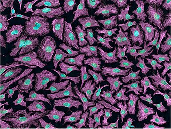
Multiphoton fluorescence image of HeLa cells with cytoskeletal microtubules (magenta) and DNA (cyan). Nikon RTS2000MP custom laser scanning microscope.
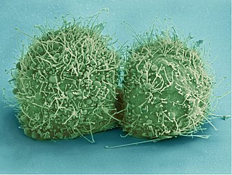
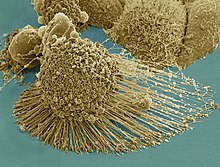
Scanning electron micrograph of just-divided HeLa cells. Zeiss Merlin HR-SEM.

HeLa cells stained with Hoechst 33258

HeLa cells grown in culture and stained with antibody to tubulin (green), antibody to Ki-67 (red) and the blue DNA binding dye DAPI. The tubulin antibody shows the distribution of microtubules and the Ki-67 antibody is expressed in cells about to divide. Preparation, antibodies and image courtesy of EnCor Biotechnology.

A volumetric surface render (red) of the nuclear envelope of one HeLa cell. The cell was observed in 300 slices of electron microscopy, the nuclear envelope was automatically segmented and rendered. One vertical and one horizontal slice are added for reference.
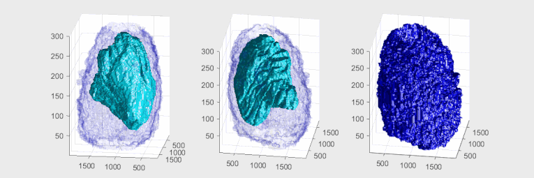
Plasma Membrane and Nuclear Envelope of one Hela Cell displayed as a volumetric surface rendering. Left and centre show the plasma membrane in blue colour with transparency and the nuclear envelope with a solid cyan colour. Right show the plasma membrane without transparency and the same angle of view as the centre. The membranes have been segmented from data acquired with Electron Microscopy.


In media[]
The 1997 documentary by Adam Curtis explains the history of HeLa and its implications in medicine and society.[59]
HeLa was the subject of a 2010 book by Rebecca Skloot, The Immortal Life of Henrietta Lacks, investigating the historical context of the cell line and how the Lacks family was involved in its use.[11] A 2017 HBO film, The Immortal Life of Henrietta Lacks was based on the book The film stars Oprah Winfrey, , Rocky Carroll and Renee Elise Goldsberry as Henrietta Lacks. Author Rebecca Skloot also appears as a character in the film, portrayed by Rose Byrne.[60]
A 2010 episode of Law & Order "Immortal" was heavily based on the story of Henrietta Lacks and the HeLa line, using the fictional "NaRo" cells as a stand-in.[61]
The story of how the HeLa line came to be was also the subject of a 2010 episode of the podcast Radiolab.[62]
A 2019 novelette by N. K. Jemisin titled Emergency Skin involves a future agent arriving on the abandoned Earth in search of HeLa culture.[63]
See also[]
- Clonally transmissible cancer
- Moore v. Regents of the University of California, case that set precedent for discarded tissue
- List of contaminated cell lines
- WI-38
References[]
- ^ Rahbari R, Sheahan T, Modes V, Collier P, Macfarlane C, Badge RM (2009). "A novel L1 retrotransposon marker for HeLa cell line identification". BioTechniques. 46 (4): 277–284. doi:10.2144/000113089. PMC 2696096. PMID 19450234.
- ^ Scherer, W.F.; Syverton, J.T.; Gey, G.O. (1953). "Studies on the propagation in vitro of poliomyelitis viruses. IV. Viral multiplication in a stable strain of human malignant epithelial cells (strain HeLa) derived from an epidermoid carcinoma of the cervix". Journal of Experimental Medicine. 97 (5): 695–710. doi:10.1084/jem.97.5.695. PMC 2136303. PMID 13052828.
- ^ "Johns Hopkins Magazine -- April 2000". pages.jh.edu.
- ^ Jump up to: a b Capes-Davis A, Theodosopoulos G, Atkin I, Drexler HG, Kohara A, MacLeod RA, Masters JR, Nakamura Y, Reid YA, Reddel RR, Freshney RI (2010). "Check your cultures! A list of cross-contaminated or misidentified cell lines". Int. J. Cancer. 127 (1): 1–8. doi:10.1002/ijc.25242. PMID 20143388. S2CID 2929020.
- ^ Jump up to: a b Batts DW (2010-05-10). "Cancer cells killed Henrietta Lacks – then made her immortal". The Virginian-Pilot. pp. 1, 12–14. Archived from the original on 2016-11-25. Retrieved 2020-05-08.
- ^ Ron Claiborne; Sydney Wright, IV (2010-01-31). "How One Woman's Cells Changed Medicine". ABC World News. Retrieved 2012-08-19.
- ^ McKie, Robin; Editor, Science (2010-04-03). "Henrietta Lacks's cells were priceless, but her family can't afford a hospital". The Guardian. London. Retrieved 18 July 2017.CS1 maint: extra text: authors list (link)
- ^ Jump up to: a b c d Lucey, Brendan P.; Nelson-Rees, Walter A.; Hutchins, Grover M. (2009-10-21). "Henrietta Lacks, HeLa Cells, and Cell Culture Contamination". Archives of Pathology & Laboratory Medicine Online. 133 (9): 1463–7. doi:10.5858/133.9.1463. PMID 19722756.
- ^ Butanis, Benjamin. "The Legacy of Henrietta Lacks". www.hopkinsmedicine.org. Retrieved 2020-05-07.
- ^ Washington, Harriet "Henrietta Lacks: An Unsung Hero", Emerge Magazine, October 1994
- ^ Jump up to: a b Zielinski, Sarah (2010-01-02). "Cracking the code of the human genome – Henrietta Lacks' 'immortal' cells". Smithsonian. Retrieved 2017-05-27.
- ^ Ritter, Malcolm (August 7, 2013). "Feds, family reach deal on use of DNA information". Seattle Times. Retrieved 2017-05-27.
- ^ del Carpio, Alexandra (April 27, 2014). "The Good, the Bad, and the HeLa". Berkeley Science Review. Retrieved 2017-05-27.
- ^ Sharrer T (2006). ""HeLa" Herself". The Scientist. 20 (7): 22.
- ^ Day, Jo Ann. "Upholding the Highest Bioethical Standards | Johns Hopkins Medicine". www.hopkinsmedicine.org. Retrieved 2020-04-15.
- ^ Jump up to: a b c Skloot, Rebecca (2010). The Immortal Life of Henrietta Lacks. New York: Crown/Random House. ISBN 978-1-4000-5217-2.
- ^ "HeLa Cell Line Origins, Contamination, Controversy, and Cytogenetics". Retrieved 2020-04-15.
- ^ Puck, T.T.; Marcus, P.I. (1955). "A rapid method for viable cell titration and clone production with Hela cells in tissue culture: The use of X-irradiated cells to supply conditioning factors". Proceedings of the National Academy of Sciences of the United States of America (published July 15, 1955). 41 (7): 432–437. Bibcode:1955PNAS...41..432P. doi:10.1073/pnas.41.7.432. PMC 528114. PMID 16589695.
- ^ Smith, Van (April 17, 2002). "Wonder Woman: The Life, Death, and Life After Death of Henrietta Lacks, Unwitting Heroine of Modern Medical Science". Baltimore City Paper. Archived from the original on August 14, 2004. Retrieved 2 March 2017.
- ^ Scherer, W.F.; Syverton, J.T.; Gey, G.O. (1953). "Studies on the Propagation in Vitro of Poliomyelitis Viruses: Iv. Viral Multiplication in a Stable Strain of Human Malignant Epithelial Cells (Strain HeLa) Derived from an Epidermoid Carcinoma of the Cervix". Journal of Experimental Medicine. 97 (5): 695–710. doi:10.1084/jem.97.5.695. PMC 2136303. PMID 13052828.
- ^ Jump up to: a b Masters, John R. (2002). "HeLa cells 50 years on: The good, the bad and the ugly". Nature Reviews Cancer. 2 (4): 315–319. doi:10.1038/nrc775. PMID 12001993. S2CID 991019.
- ^ Turner, Timothy (2012). "Development of the Polio Vaccine: A Historical Perspective of Tuskegee University's Role in Mass Production and Distribution of HeLa Cells". Journal of Health Care for the Poor and Underserved. 23 (4a): 5–10. doi:10.1353/hpu.2012.0151. PMC 4458465. PMID 23124495.
- ^ Brownlee, K. A. (1955). "Statistics of the 1954 Polio Vaccine Trials*". Journal of the American Statistical Association. 50 (272): 1005–1013. doi:10.1080/01621459.1955.10501286.
- ^ Parker, J; Murphy W; Wang D; O'Brien S; Parrish C (2001). "Canine and feline parvoviruses can use human or feline transferrin receptors to bind, enter, and infect cells". Journal of Virology. 75 (8): 3896–3902. doi:10.1128/JVI.75.8.3896-3902.2001. PMC 114880. PMID 11264378.
- ^ Acrani, G.O.; Gomes, R.; Proença-Módena, J.L.; da Silva, A.F.; Carminati, P.O.; Silva, M.L.; Santos, R.I.; Arruda, E. (2010). "Apoptosis induced by Oropouche virus infection in HeLa cells is dependent on virus protein expression". Virus Research. 149 (1): 56–63. doi:10.1016/j.virusres.2009.12.013. PMID 20080135.
- ^ Hou, S.Y.; Wu, S.; Chiang, C. (2002). "Transcriptional activity among high and low risk human papillomavirus E2 proteins correlates with E2 DNA binding". The Journal of Biological Chemistry. 277 (47): 45619–45629. doi:10.1074/jbc.M206829200. PMID 12239214. S2CID 9203953.
- ^ Jump up to: a b del Puerto, H.L.; Martins, A.S.; Milsted, A.; Souza-Fagundes, E.M.; Braz, G.F.; Hissa, B.; Andrade, L.O.; Alves, F.; Rajão, D.S.; Leite, R.C.; Vasconcelos, A.C. (2011). "Canine distemper virus induces apoptosis in cervical tumor derived cell lines". Virol. J. 8 (1): 334. doi:10.1186/1743-422X-8-334. PMC 3141686. PMID 21718481.
- ^ Jump up to: a b c "5 Contributions HeLa Cells Have Made to Science". Cell Science from Technology Networks. Retrieved 2020-03-25.
- ^ Mondor, Isabelle; Ugolini, Sophie; Sattentau, Quentin J. (1998-05-01). "Human Immunodeficiency Virus Type 1 Attachment to HeLa CD4 Cells Is CD4 Independent and gp120 Dependent and Requires Cell Surface Heparans". Journal of Virology. 72 (5): 3623–3634. doi:10.1128/jvi.72.5.3623-3634.1998. ISSN 1098-5514. PMC 109583. PMID 9557643.
- ^ Wechsler, Steven L.; Rustigian, Robert; Stallcup, Kathryn C.; Byers, Karen B.; Winston, Stuart H.; Fields, Bernard N. (1979). "Measles Virus-Specified Polypeptide Synthesis in Two Persistently Infected HeLa Cell Lines". Journal of Virology. 31 (3): 677–684. doi:10.1128/jvi.31.3.677-684.1979. ISSN 0022-538X. PMC 353496. PMID 513191.
- ^ Li, Li; Collins, Natalie D.; Widen, Steven G.; Davis, Emily H.; Kaiser, Jaclyn A.; White, Mellodee M.; Greenberg, M. Banks; Barrett, Alan D. T.; Bourne, Nigel; Sarathy, Vanessa V. (2019-08-20). "Attenuation of Zika Virus by Passage in Human HeLa Cells". Vaccines. 7 (3): 93. doi:10.3390/vaccines7030093. ISSN 2076-393X. PMC 6789458. PMID 31434319.
- ^ Pamela Bulzomi; Paola Galluzzo; Alessandro Bolli; Stefano Leone; Filippo Acconcia; Maria Marino (2012). "The pro-apoptotic effect of quercetin in cancer cell lines requires ERβ-dependent signals". Journal of Cellular Physiology. 227 (5): 1891–1898. doi:10.1002/jcp.22917. PMID 21732360. S2CID 24034074.
- ^ Tan X, Luo S, Wang D, Su Y, Cheng T, Shi C (2011). "A NIR heptamethine Dye with intrinsic cancer targeting, imaging and photosynthesizing properties". Journal of Biomaterials China. 33 (7): 2230–2239. doi:10.1016/j.biomaterials.2011.11.081. PMID 22182749.
- ^ Pene, F.; Courtine, E.; Cariou, A.; Mira, J.P. (2009). "Toward theranostics". Crit Care Med. 37 (1 Suppl): S50–S58. doi:10.1097/CCM.0b013e3181921349. PMID 19104225. S2CID 37043095.
- ^ Briiuner., Thomas; Dieter F. Hulser (1990). "Tumor Cell Invasion and Gap Junctional Communication" (PDF). Invasion Metastasis. 10: 31–34. Retrieved 3 April 2012.
- ^ Xie, Z.; Wroblewska, L.; Prochazka, L.; Weiss, R.; Benenson, Y. (2011). "Multi-Input RNAi-Based Logic Circuit for Identification of Specific Cancer Cells" (PDF). Science. 333 (6047): 1307–1311. Bibcode:2011Sci...333.1307X. doi:10.1126/science.1205527. PMID 21885784. S2CID 13743291.
- ^ Arjomandnejad, M; et al. (2014). "HeLa cell line xenograft tumor as a suitable cervical cancer model: growth kinetic characterization and immunohistochemistry array" (PDF). Archives of Iranian Medicine. 17 (4): 273–277. PMID 24724604. S2CID 25652255. Archived from the original (PDF) on 2018-04-25.
- ^ Davies, Preston. "Significant Research Advances Enabled by HeLa Cells". Office of Science Policy. Retrieved 2020-03-25.
- ^ The Nobel Prize in Physiology or Medicine 2009 on nobelprize.org
- ^ Ivanković M, Cukusić A, Gotić I, Skrobot N, Matijasić M, Polancec D, Rubelj I (2007). "Telomerase activity in HeLa cervical carcinoma cell line proliferation". Biogerontology. 8 (2): 163–72. doi:10.1007/s10522-006-9043-9. PMID 16955216. S2CID 9390790.
- ^ Jump up to: a b c Macville M, Schröck E, Padilla-Nash H, Keck C, Ghadimi BM, Zimonjic D, Popescu N, Ried T (1999). "Comprehensive and definitive molecular cytogenetic characterization of HeLa cells by spectral karyotyping". Cancer Res. 59 (1): 141–50. PMID 9892199.
- ^ Landry JJ, Pyl PT, Rausch T, Zichner T, Tekkedil MM, Stütz AM, Jauch A, Aiyar RS, Pau G, Delhomme N, Gagneur J, Korbel JO, Huber W, Steinmetz LM (2013). "The genomic and transcriptomic landscape of a HeLa cell line". G3: Genes, Genomes, Genetics. 3 (8): 1213–24. doi:10.1534/g3.113.005777. PMC 3737162. PMID 23550136.
- ^ Bottomley, R.H.; Trainer, A.L.; Griffin, M.J. (1969). "Enzymatic and chromosomal characterization of HeLa variants". J. Cell Biol. 41 (3): 806–15. doi:10.1083/jcb.41.3.806. PMC 2107821. PMID 5768876.
- ^ Andrew Adey; Joshua N. Burton; Jacob O. Kitzman; Joseph B. Hiatt; Alexandra P. Lewis; Beth K. Martin; Ruolan Qiu; Choli Lee; Jay Shendure (2013). "The haplotype-resolved genome and epigenome of the aneuploid HeLa cancer cell line". Nature (published August 8, 2013). 500 (7461): 207–211. Bibcode:2013Natur.500..207A. doi:10.1038/nature12064. PMC 3740412. PMID 23925245.
- ^ Landry JJ, Pyl PT, Rausch T, Zichner T, Tekkedil MM, Stütz AM, Jauch A, Aiyar RS, Pau G, Delhomme N, Gagneur J, Korbel JO, Huber W, Steinmetz LM (2013). "The genomic and transcriptomic landscape of a HeLa cell line". G3: Genes, Genomes, Genetics. 3 (8): 1213–24. doi:10.1534/g3.113.005777. PMC 3737162. PMID 23550136.
- ^ Callaway, Ewen (March 15, 2013). "Most popular human cell in science gets sequenced". Nature. doi:10.1038/nature.2013.12609. S2CID 87549985. Retrieved 8 August 2013.
- ^ Jump up to: a b Callaway, Ewen (March 27, 2013). "HeLa publication brews bioethical storm". Nature. doi:10.1038/nature.2013.12689. S2CID 88020977. Retrieved 8 August 2013.
- ^ Callaway, Ewen (August 7, 2013). "Deal done over HeLa cell line". Nature. 500 (7461): 132–133. Bibcode:2013Natur.500..132C. doi:10.1038/500132a. PMID 23925220.
- ^ Jump up to: a b "NIH, Lacks family reach understanding to share genomic data of HeLa cells". The National Institutes of Health. August 7, 2013. Retrieved 8 August 2013.
- ^ Callaway, Ewen (August 7, 2013). "NIH director explains HeLa agreement". Nature. doi:10.1038/nature.2013.13521. S2CID 190871214.
- ^ Gold, Michael, 1953- (1986). A conspiracy of cells : one woman's immortal legacy and the medical scandal it caused. State University of New York Press. ISBN 0-88706-099-4. OCLC 12805138.CS1 maint: multiple names: authors list (link)
- ^ Wang H, Huang S, Shou J, Su EW, Onyia JE, Liao B, Li S (2006). "Comparative analysis and integrative classification of NCI60 cell lines and primary tumors using gene expression profiling data". BMC Genomics. 7 (1): 166. doi:10.1186/1471-2164-7-166. PMC 1525183. PMID 16817967.
- ^ Nardone, R.M. (2007). "Eradication of cross-contaminated cell lines: A call for action" (PDF). Cell Biology and Toxicology. 23 (6): 367–372. CiteSeerX 10.1.1.432.8581. doi:10.1007/s10565-007-9019-9. PMID 17522957. S2CID 21077969. Archived from the original (PDF) on 2008-12-17. Retrieved 2007-12-04.
- ^ "ATCC® Standards Development Organization: The International Cell Line Authentication Committee (ICLAC)". Standards.atcc.org. Retrieved 2013-06-27.
- ^ Maiorana VC, Van Valen LM (7 February 1991). "Hela, a new microbial species" (PDF). Evolutionary Theory. 10 (2): 71–4. Archived (PDF) from the original on 29 March 2018. Lay summary.
- ^ Duesberg, P; Mandrioli, D; McCormack, A; Nicholson, JM (2011). "Is carcinogenesis a form of speciation?". Cell Cycle. Georgetown, Texas. 10 (13): 2100–2114. doi:10.4161/cc.10.13.16352. PMID 21666415.
- ^ van Valen, L.M.; Maiorana, V.C. (1991). "HeLa, a new microbial species". Evolutionary Theory & Review. 10: 71–74. ISSN 1528-2619.
- ^ Cohan, FM (2002). "What are bacterial species?". Annu. Rev. Microbiol. 56 (1): 457–487. doi:10.1146/annurev.micro.56.012302.160634. PMID 12142474.
- ^ "Modern Times: The Way of All Flesh - BBC Two England - 19 March 1997". The Radio Times (3815): 92. 1997-03-13.
- ^ "About The Immortal Life of Henrietta Lacks". Rebecca Skloot. Retrieved 24 April 2018.
- ^ Thomas, June (2010-05-19). "Ripped From Which Headline? "Immortal"". Slate Magazine. Retrieved 2021-08-03.
- ^ "Henrietta's Tumor".
- ^ "Emergency Skin".
Further reading[]
- Hannah Landecker (2000). "Immortality, In Vitro: A History of the HeLa Cell Line". In Brodwin, Paul (ed.). Biotechnology and culture: bodies, anxieties, ethics. Bloomington: Indiana University Press. pp. 53–74. ISBN 978-0-253-21428-7.
- Rebecca Skloot (2010). The Immortal Life of Henrietta Lacks.
External links[]
| Wikimedia Commons has media related to HeLa cells. |
- HeLa (CCL-2 Cells) in the ATCC database
- HeLa Cells at the US National Library of Medicine Medical Subject Headings (MeSH)
- HeLa Transfection and Selection Data for HeLa Cells
- Rebecca Skloot, The Immortal Life of Henrietta Lacks book website with additional features (photo/video/audio)
- The Henrietta Lacks Foundation, a foundation established to, among other things, help provide scholarship funds and health insurance to Henrietta Lacks's family.
- Rebecca Skloot, Cells That Save Lives are a Mother's Legacy, New York Times
- "Wonder Woman: The Life, Death, and Life After Death of Henrietta Lacks, Unwitting Heroine of Modern Medical Science" by Van Smith
- "What's Left of Henrietta Lacks?" by Anne Enright
- "Culturing Life: How Cells Became Technologies" a book by Hannah Landecker about HeLa and the history of tissue culture.
- Discussion about the taxonomic effect of creating the new taxon Helacyton.
- Cell Centered Database – HeLa cell
- Audio Interview with Rebecca Skloot about her book "The Immortal Life of Henrietta Lacks"
- Cellosaurus entry for HeLa
- The Legacy of Henrietta Lacks
- Human cell lines
- Bioethics
- Johns Hopkins Hospital
- Cellular senescence
- 1951 in biotechnology