Lamina affixa
| Lamina affixa | |
|---|---|

| |
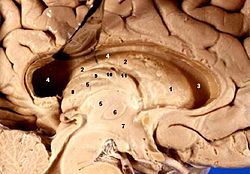 Human brain left dissected midsagittal view (Lamina affixa is #10) | |
| Details | |
| Identifiers | |
| Latin | lamina affixa |
| TA98 | A14.1.09.276 |
| TA2 | 5650 |
| FMA | 83709 |
| Anatomical terms of neuroanatomy | |
Lamina affixa is a layer of epithelium growing on the surface of the thalamus and forming the floor of the central part of lateral ventricle, on whose medial margin is attached the choroid plexus of the lateral ventricle; it covers the superior thalamostriate vein and the superior choroid vein. The torn edge of this plexus is called the tela choroidea.[1]
On the surface of the terminal vein is a narrow white band, named the lamina affixa.
GDF-15/MIC-1 has been observed in lamina affixa cells.[2]
References[]
![]() This article incorporates text in the public domain from page 838 of the 20th edition of Gray's Anatomy (1918)
This article incorporates text in the public domain from page 838 of the 20th edition of Gray's Anatomy (1918)
- ^ Alberts, Daniel; et al. (2012). Dorland's illustrated medical dictionary (32nd ed.). Philadelphia, PA: Saunders/Elsevier. p. 1878. ISBN 978-1-4160-6257-8.
- ^ Schober A, Böttner M, Strelau J, et al. (October 2001). "Expression of growth differentiation factor-15/ macrophage inhibitory cytokine-1 (GDF-15/MIC-1) in the perinatal, adult, and injured rat brain". J. Comp. Neurol. 439 (1): 32–45. doi:10.1002/cne.1333. PMID 11579380. S2CID 40302340.
External links[]
| Authority control: Scientific databases |
|---|
This neuroanatomy article is a stub. You can help Wikipedia by . |
Categories:
- Wikipedia articles incorporating text from the 20th edition of Gray's Anatomy (1918)
- Thalamus
- Neuroanatomy stubs