Necrotizing sialometaplasia
| Necrotizing sialometaplasia | |
|---|---|
 | |
| Specialty | ENT surgery |
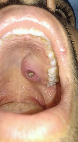
Necrotizing sialometaplasia (NS) is a benign, ulcerative lesion, usually located towards the back of the hard palate. It is thought to be caused by ischemic necrosis (death of tissue due to lack of blood supply) of minor salivary glands in response to trauma. Often painless, the condition is self-limiting and should heal in 6–10 weeks.
Although entirely benign and requiring no treatment, due to its similar appearance to oral cancer, it is sometimes misdiagnosed as malignant. Therefore, it is considered an important condition, despite its rarity.
Signs and symptoms[]
The condition most commonly is located at the junction of the hard and soft palate.[1] However, the condition may arise anywhere minor salivary glands are located.[nb 1] It has also been occasionally reported to involve the major salivary glands.[2][3] It may be present only on one side, or both sides.[1] The lesion typically is 1–4 cm in diameter.[4]
Initially, the lesion is a tender, erythematous (red) swelling. Later, in the ulcerated stage, the overlying mucosa breaks down to leave a deep, well-circumscribed ulcer which is yellow-gray in color and has a lobular base.[1]
There is usually only minor pain,[1] and the condition is often entirely painless. There may be prodromal symptoms similar to flu before the appearance of the lesion.[4]
Causes[]
The exact cause of the condition is unknown.[4][5] There is most evidence to support vascular infarction and ischemic necrosis of salivary gland lobules as a mechanism for the condition.[6] Experimentally, local anaesthetic injections and tying of the arteries is reported to trigger the development of tissue changes similar to NS in lab rats.[6] Factors which are thought to cause this ischemia are listed below, however sometimes there is no evident predisposing factor or initiating event.[6]
- Trauma[4] e.g. during intubation,[6] or surgical procedures[6]
- Local anesthetic injection[4]
- Smoking[4]
- Alcohol[6]
- Diabetes mellitus[4]
- Vascular disease,[4] (e.g. arteriosclerosis)[5]
- Pressure from a dental prosthesis[4]
- Allergy[5]
- Bulimia[2]
- Infection[6]
- Ionizing radiation[6]
Diagnosis[]
Differentiation between this and SCC would be based on a history of recent trauma or dental treatment in the area.
Immunohistochemistry may aid the diagnosis. If the lesion is NS, there will be focal to absent immunoreactivity for p53, low immunoreactivity for MIB1 (Ki-67), and the presence of /p63- and calponin-positive myoepithelial cells.[2]
Treatment[]
No surgery is required.[4]
Prognosis[]
Healing is prolonged, and usually takes 6–10 weeks.[1] The ulcer heals by secondary intention.[7]
Epidemiology[]
The condition is rare.[8][9] The typical age range of those affected by the condition is about 23–66 years of age.[4] It usually occurs in smokers.[9] The male to female ratio has been reported as 1.95:1,[5] and 2.31:1.[10]
History[]
NS was first reported by Abrams et al. in 1973.[11][6]
Notes[]
References[]
- ^ a b c d e Regezi JA; Scuibba JJ; Jordan RCK (2012). Oral pathology : clinical pathologic correlations (6th ed.). St. Louis, Mo.: Elsevier/Saunders. p. 191. ISBN 978-1-4557-0262-6.
- ^ a b c Carlson, DL (May 2009). "Necrotizing sialometaplasia: a practical approach to the diagnosis". Archives of Pathology & Laboratory Medicine. 133 (5): 692–8. doi:10.1043/1543-2165-133.5.692. PMID 19415943.
- ^ Tsuji, T; Nishide, Y; Nakano, H; Kida, K; Satoh, K (2014). "Imaging findings of necrotizing sialometaplasia of the parotid gland: case report and literature review". Dentomaxillofacial Radiology. 43 (6): 20140127. doi:10.1259/dmfr.20140127. PMC 4141672. PMID 24850145.
- ^ a b c d e f g h i j k Hupp JR; Tucker MR; Ellis E (19 March 2013). Contemporary Oral and Maxillofacial Surgery (6th ed.). Elsevier Health Sciences. pp. 412–414. ISBN 978-0-323-22687-5.
- ^ a b c d Schmidt-Westhausen, A; Philipsen, HP; Reichart, PA (1991). "[Necrotizing sialometaplasia of the palate. Literature report of 3 new cases]". Deutsche Zeitschrift für Mund-, Kiefer- und Gesichts-Chirurgie. 15 (1): 30–4. PMID 1814663.
- ^ a b c d e f g h i Barnes L (2008). Surgical pathology of the head and neck (3rd ed.). New York: Informa Healthcare. pp. 491–493. ISBN 9780849390234.
- ^ Imbery, TA; Edwards, PA (July 1996). "Necrotizing sialometaplasia: literature review and case reports". Journal of the American Dental Association. 127 (7): 1087–92. doi:10.14219/jada.archive.1996.0334. PMID 8754467.
- ^ Janner, SF; Suter, VG; Altermatt, HJ; Reichart, PA; Bornstein, MM (May 2014). "Bilateral necrotizing sialometaplasia of the hard palate in a patient with bulimia: a case report and review of the literature". Quintessence International. 45 (5): 431–7. doi:10.3290/j.qi.a31543. PMID 24634907.
- ^ a b Scully C (2013). Oral and maxillofacial medicine: the basis of diagnosis and treatment (3rd ed.). Edinburgh: Churchill Livingstone. p. 405. ISBN 9780702049484.
- ^ Jainkittivong, A; Sookasam, M; Philipsen, HP (1989). "Necrotizing sialometaplasia: review of 127 cases". The Journal of the Dental Association of Thailand. 39 (1): 11–6. PMID 2699611.
- ^ Abrams, AM; Melrose, RJ; Howell, FV (July 1973). "Necrotizing sialometaplasia. A disease simulating malignancy". Cancer. 32 (1): 130–5. doi:10.1002/1097-0142(197307)32:1<130::aid-cncr2820320118>3.0.co;2-8. PMID 4716764.
External links[]
- Salivary gland pathology