40S ribosomal protein S27a is a protein that in humans is encoded by the RPS27Agene.[5][6]
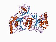
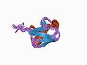
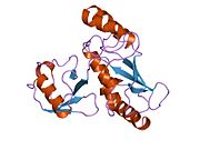
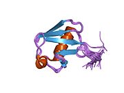
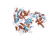
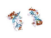
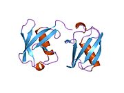
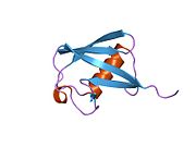
Ubiquitin, a highly conserved protein that has a major role in targeting cellular proteins for degradation by the 26S proteosome, is synthesized as a precursor protein consisting of either polyubiquitin chains or a single ubiquitin fused to an unrelated protein. This gene encodes a fusion protein consisting of ubiquitin at the N terminus and ribosomal protein S27a at the C terminus. When expressed in yeast, the protein is post-translationally processed, generating free ubiquitin monomer and ribosomal protein S27a. Ribosomal protein S27a is a component of the 40S subunit of the ribosome and belongs to the S27AE family of ribosomal proteins. It contains C4-type zinc finger domains and is located in the cytoplasm. Pseudogenes derived from this gene are present in the genome. As with ribosomal protein S27a, ribosomal protein L40 is also synthesized as a fusion protein with ubiquitin; similarly, ribosomal protein S30 is synthesized as a fusion protein with the ubiquitin-like protein fubi.[6]
Pancré V, Pierce RJ, Fournier F, et al. (1991). "Effect of ubiquitin on platelet functions: possible identity with platelet activity suppressive lymphokine (PASL)". Eur. J. Immunol. 21 (11): 2735–41. doi:10.1002/eji.1830211113. PMID1657614. S2CID23901646.
Kanayama H, Tanaka K, Aki M, et al. (1992). "Changes in expressions of proteasome and ubiquitin genes in human renal cancer cells". Cancer Res. 51 (24): 6677–85. PMID1660345.
Redman KL, Rechsteiner M (1989). "Identification of the long ubiquitin extension as ribosomal protein S27a". Nature. 338 (6214): 438–40. Bibcode:1989Natur.338..438R. doi:10.1038/338438a0. PMID2538756. S2CID4360107.
Maruyama K, Sugano S (1994). "Oligo-capping: a simple method to replace the cap structure of eukaryotic mRNAs with oligoribonucleotides". Gene. 138 (1–2): 171–4. doi:10.1016/0378-1119(94)90802-8. PMID8125298.
Suzuki Y, Yoshitomo-Nakagawa K, Maruyama K, et al. (1997). "Construction and characterization of a full length-enriched and a 5'-end-enriched cDNA library". Gene. 200 (1–2): 149–56. doi:10.1016/S0378-1119(97)00411-3. PMID9373149.
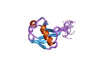
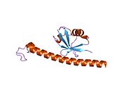
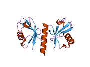
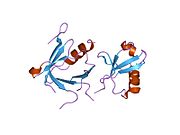
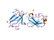
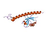
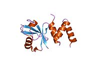
Bolton D, Evans PA, Stott K, Broadhurst RW (2002). "Structure and properties of a dimeric N-terminal fragment of human ubiquitin". J. Mol. Biol. 314 (4): 773–87. doi:10.1006/jmbi.2001.5181. PMID11733996.
1aar: STRUCTURE OF A DIUBIQUITIN CONJUGATE AND A MODEL FOR INTERACTION WITH UBIQUITIN CONJUGATING ENZYME (E2)
1cmx: STRUCTURAL BASIS FOR THE SPECIFICITY OF UBIQUITIN C-TERMINAL HYDROLASES
1d3z: UBIQUITIN NMR STRUCTURE
1f9j: STRUCTURE OF A NEW CRYSTAL FORM OF TETRAUBIQUITIN
1fxt: STRUCTURE OF A CONJUGATING ENZYME-UBIQUITIN THIOLESTER COMPLEX
1g6j: STRUCTURE OF RECOMBINANT HUMAN UBIQUITIN IN AOT REVERSE MICELLES
1gjz: SOLUTION STRUCTURE OF A DIMERIC N-TERMINAL FRAGMENT OF HUMAN UBIQUITIN
1nbf: Crystal structure of a UBP-family deubiquitinating enzyme in isolation and in complex with ubiquitin aldehyde
1ogw: SYNTHETIC UBIQUITIN WITH FLUORO-LEU AT 50 AND 67
1otr: Solution Structure of a CUE-Ubiquitin Complex
1p3q: Mechanism of Ubiquitin Recognition by the CUE Domain of VPS9
1q0w: Solution structure of Vps27 amino-terminal UIM-ubiquitin complex
1q5w: Ubiquitin Recognition by Npl4 Zinc-Fingers
1s1q: TSG101(UEV) domain in complex with Ubiquitin
1sif: Crystal structure of a multiple hydrophobic core mutant of ubiquitin
1tbe: STRUCTURE OF TETRAUBIQUITIN SHOWS HOW MULTIUBIQUITIN CHAINS CAN BE FORMED
1ubi: SYNTHETIC STRUCTURAL AND BIOLOGICAL STUDIES OF THE UBIQUITIN SYSTEM. PART 1
1ubq: STRUCTURE OF UBIQUITIN REFINED AT 1.8 ANGSTROMS RESOLUTION
1ud7: SOLUTION STRUCTURE OF THE DESIGNED HYDROPHOBIC CORE MUTANT OF UBIQUITIN, 1D7
1uzx: A COMPLEX OF THE VPS23 UEV WITH UBIQUITIN
1v80: Solution structures of ubiquitin at 30 bar and 3 kbar
1v81: Solution structures of ubiquitin at 30 bar and 3 kbar
1wr1: The complex structure of Dsk2p UBA with ubiquitin
1wr6: Crystal structure of GGA3 GAT domain in complex with ubiquitin
1wrd: Crystal structure of Tom1 GAT domain in complex with ubiquitin
1xd3: Crystal structure of UCHL3-UbVME complex
1xqq: Simultaneous determination of protein structure and dynamics
1yd8: COMPLEX OF HUMAN GGA3 GAT DOMAIN AND UBIQUITIN
1yiw: X-ray Crystal Structure of a Chemically Synthesized Ubiquitin
1yj1: X-ray Crystal Structure of a Chemically Synthesized [D-Gln35]Ubiquitin
1yx5: Solution Structure of S5a UIM-1/Ubiquitin Complex
1yx6: Solution Structure of S5a UIM-2/Ubiquitin Complex
1zgu: Solution structure of the human Mms2-Ubiquitin complex
2ayo: Structure of USP14 bound to ubquitin aldehyde
2bgf: NMR STRUCTURE OF LYS48-LINKED DI-UBIQUITIN USING CHEMICAL SHIFT PERTURBATION DATA TOGETHER WITH RDCS AND 15N-RELAXATION DATA
2c7m: HUMAN RABEX-5 RESIDUES 1-74 IN COMPLEX WITH UBIQUITIN
2c7n: HUMAN RABEX-5 RESIDUES 1-74 IN COMPLEX WITH UBIQUITIN
2d3g: Double sided ubiquitin binding of Hrs-UIM
2den: Solution Structure of the Ubiquitin-Associated Domain of Human BMSC-UbP and its Complex with Ubiquitin
2dx5: The complex structure between the mouse EAP45-GLUE domain and ubiquitin
2fcm: X-ray Crystal Structure of a Chemically Synthesized [D-Gln35]Ubiquitin with a Cubic Space Group
2fcn: X-ray Crystal Structure of a Chemically Synthesized [D-Val35]Ubiquitin with a Cubic Space Group
2fcq: X-ray Crystal Structure of a Chemically Synthesized Ubiquitin with a Cubic Space Group
2fcs: X-ray Crystal Structure of a Chemically Synthesized [L-Gln35]Ubiquitin with a Cubic Space Group
2fid: Crystal Structure of a Bovine Rabex-5 fragment complexed with ubiquitin
2fif: Crystal Structure of a Bovine Rabex-5 fragment complexed with ubiquitin
2fuh: Solution Structure of the UbcH5c/Ub Non-covalent Complex
2g3q: Solution Structure of Ede1 UBA-ubiquitin complex
2g45: Co-crystal structure of znf ubp domain from the deubiquitinating enzyme isopeptidase T (isot) in complex with ubiquitin
2gbj: Crystal Structure of the 9-10 8 Glycine Insertion Mutant of Ubiquitin.
2gbk: Crystal Structure of the 9-10 MoaD Insertion Mutant of Ubiquitin
2gbm: Crystal Structure of the 35-36 8 Glycine Insertion Mutant of Ubiquitin
2gbn: Crystal Structure of the 35-36 8 Glycine Insertion Mutant of Ubiquitin
2gbr: Crystal Structure of the 35-36 MoaD Insertion Mutant of Ubiquitin
2gmi: Mms2/Ubc13~Ubiquitin
2hd5: USP2 in complex with ubiquitin
2hth: Structural basis for ubiquitin recognition by the human EAP45/ESCRT-II GLUE domain
2ibi: Covalent Ubiquitin-USP2 Complex
2j7q: CRYSTAL STRUCTURE OF THE UBIQUITIN-SPECIFIC PROTEASE ENCODED BY MURINE CYTOMEGALOVIRUS TEGUMENT PROTEIN M48 IN COMPLEX WITH A UBQUITIN-BASED SUICIDE SUBSTRATE
2nr2: The MUMO (minimal under-restraining minimal over-restraining) method for the determination of native states ensembles of proteins
2o6v: Crystal structure and solution NMR studies of Lys48-linked tetraubiquitin at neutral pH
2oob: crystal structure of the UBA domain from Cbl-b ubiquitin ligase in complex with ubiquitin