Radiodonta
| Radiodonta | |
|---|---|
| |
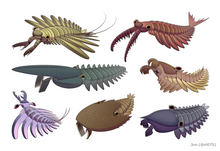
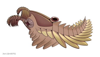
| Left to right, top to bottom: Amplectobelua symbrachiata, Anomalocaris canadensis, Aegirocassis benmoulai, Peytoia nathorsti, Lyrarapax unguispinus, Cambroraster falcatus, and Hurdia victoria | |
| Scientific classification | |
| Kingdom: | |
| Phylum: | |
| Class: | |
| Order: | †Radiodonta
Collins, 1996 |
| Families | |
| |
Radiodonta is an order of stem-group arthropods that was successful worldwide during the Cambrian period. They may be referred to as radiodonts,[1][2][3] radiodontans,[4][5] radiodontids,[6] anomalocarids,[7] or anomalocaridids,[8][9][10] although the latter originally refer to the family Anomalocarididae, which previously included all species of this order but is now restricted to only a few species.[7] Radiodonts are distinguished by their distinctive frontal appendages, which are morphologically diverse and used for a variety of functions. Radiodonts included the earliest large predators known, but they also included sediment sifters and filter feeders. Some of the most famous species of radiodonts are the Cambrian taxa Anomalocaris canadensis, Hurdia victoria, Peytoia nathorsti, and Amplectobelua symbrachiata, the Ordovician Aegirocassis benmoulai and the Devonian Schinderhannes bartelsi.
Etymology[]
The name Radiodonta (Latin for radius "spoke of a wheel" and Greek for odoús "tooth") refers to the radial arrangement of tooth plates (oral cone) surrounding the mouth,[6] although these features are suggested to be absent in some radiodont species.[4][1]
Definition[]
The original diagnosis of order Radiodonta in 1996 is as follows:[6]
Radiodontids are bilaterally symmetrical, elongate arthropods with a nonmineralized cuticle typically most robust in the jaws and claws. The body is subdivided into two tagmata, much like the prosoma and opisthosoma of chelicerate arthropods. Typically, the front part shows no external segmentation, bears one pair of preoral claws, a pair of prominent eyes, and ventral jaws with radiating teeth. Some forms have additional rows of teeth and three or four postoral limb pairs. The trunk is metameric, typically with about 13 segments laterally developing imbricating lobes for swimming and gills for respiration, and may end in a prominent three-part tail. Some forms have gnathobasic trunk limbs.
In 2014, the clade Radiodonta was defined phylogenetically as a clade including any taxa closer to Anomalocaris canadensis than Paralithodes camtschaticus.[7] In 2019, it was redefined morphologically as animal bearing head carapace complex with central (H-) and lateral (P-) elements; outgrowths (endites) from frontal appendages bearing auxiliary spines; and reduced anterior flaps or bands of lamellae (setal blades) and strong tapering of body from anterior to posterior.[3]

Description[]

Most radiodonts were significantly larger than other Cambrian fauna, with typical body lengths varying from 30 to 50 centimeters.[2] The largest described radiodont is Ordovician Aegirocassis benmoulai, which may have grown up to two meters long.[10][2] A nearly complete specimen of a juvenile Lyrarapax unguispinus measured only 18 millimetres (0.71 in), making it among the smallest radiodont specimens known, though adults reached a length of 8 centimetres (3.1 in)[2][11] An isolated frontal appendage of a hurdiid with a length less than half that of the juvenile Lyrarapax is known, but it is not known whether this specimen pertains to an adult.[12] The largest known Cambrian radiodont was Laminacaris, although known from only by frontal appendages, had an estimated body length of up to 78.4 centimetres (30.9 in) based on Anomalocaris. Anomalocaris and Amplectobelua are also large ones, reached 37.8 centimetres (14.9 in) and 48 centimetres (19 in) (There was an estimation that Houcaris saron (Previously Anomalocaris saron) reached 56 centimetres (22 in), but specimen used for estimating the body length is no longer belongs to that species[13]); the Cambrian hurdiid Titanokorys approached it in size, with an estimated body length of approximately 50 centimetres (20 in).[2][14]
The body of a radiodont could be divided into two regions: head and trunk. The head is composed of only one body segment[15] known as the ocular somite, covered by sclerites (head carapace complex), bore arthropodized frontal appendages, ventral mouthparts (oral cone), and stalked compound eyes. The tapering trunk is composed of multiple body segments, each associated with pairs of flaps and gill-like structures (setal blades).[3]
Frontal appendage[]


The anterior structures on the head are a pair of frontal appendages which have been referred to as 'claws', 'grasping appendages', 'feeding appendages', or 'great appendages' in previous studies (the last term is no longer used since the frontal appendages are now considered non-homologous to megacheiran great appendages.[15]). They are sclerotized and arthropodized (segmented), bearing ventral endites (spines) on most of their podomeres (segmental units), and the endites may bear additional rows of auxiliary spines on their anterior and posterior margins.[16][3] The frontal appendage consists of two regions: the shaft ('peduncle' in previous studies) and the distal articulated region.[16] A triangular region covered by soft cuticle (arthrodial membrane) may occur on the ventral side between podomeres and provide flexibility.[17] Their pre-ocular and protocerebral origin suggest they are homologous to the primary antennae of Onychophora and the labrum of Euarthropoda (all arose from ocular somite), and not homologous with the chelicerae of Chelicerata nor the antennae or 'great appendages' of other arthropods, which are deutocerebral (arose from post-ocular somite).[9][15] Since the morphology of the frontal appendages, especially those of the spines, always differs between species, it is one of the most important means of species identification.[16] In fact, many radiodonts are only known from a handful of fossilized frontal appendages.[17][16]

Frontal appendages of Anomalocarididae, Amplectobeluidae, and possibly related species.

Frontal appendages of Tamisiocarididae (="Cetiocaridae").

Frontal appendages of Hurdiidae.


Oral cone[]


The mouth is on the ventral side of the head, behind the attachment point of frontal appendages and is surrounded by a ring of tooth plates, forming the mouthpart known as oral cone ('jaws' in previous studies[6]). 3 or 4 tooth plates might be enlarged, giving the oral cone a triradial (e.g. Anomalocaris) or tetraradial (e.g. Hurdiidae, Lyrarapax) appearance.[18][11] The inner margin of tooth plates have spikes facing towards the mouth opening. Additional rows of internal tooth plates may occur in some hurdiid genera.[8][3] Detail reconstruction of amplectobeluid oral cones are speculative, but they possibly did not present a typical radial arrangement.[4][1]
Head sclerites, eyes and trunk[]


Three head sclerites (head carapace complex) formed by a central H-element (anterior sclerite or head shield) and a pair of P-elements (lateral sclerites) cover the dorsal and laterovental surface of the animal's head.[3] The P-elements may connect to each other as well as the H-element by a narrow anterior extension (P-element neck or 'beak').[8][3] The head carapace complex is small and ovoid in Anomalocarididae and Amplectobeluidae,[4][3] but often enlarged in Hurdiidae.[3] The head bore two stalked compound eyes, which may have had mobility,[19] and are located between the gaps formed by the posterior regions of the H-element and P-element.[8][3]


Contrary to the original diagnosis, the division of body segments (segmental boundaries) can be visible externally[10][5][3] and no known member of Radiodonta (except the putative radiodont Cucumericrus[10][20]) is known to have pediform trunk appendages (legs).[21] The trunk has numerous body segments (somites), tapering from anterior to posterior, with the anterior 3 or 4 segments significantly constricted into a neck region.[3]

The trunk appendages were fin-like body flaps ('lateral flaps' or 'lobes' in some studies), usually one pair of ventral flaps per body segment, each slightly overlapping the one more anterior to it, but additional, non-overlapping sets of small dorsal flaps may occur in some Hurdiid species.[10] The flaps may have numerous vein-like structures known as strengthening rays.[10][5][3] The flaps on the neck region, called anterior flaps or neck flaps, are significantly reduced. In some species, jaw-like feeding appendages called gnathobase-like structures (GLSs) arose from each of the bases of their reduced neck flaps.[4][1] Numerous elongated blade-like extensions (lanceolate blades) arranged in a row, forming bands of gill-like structures known as setal blades or lamellae,[3] covered the dorsal surface of each body segment.[10] At least in Aegirocassis, each of the lanceolate blades are covered in wrinkles.[10]
The ventral flaps may be homologous to the endopod of the biramous limbs of euarthropods and lobopodous limbs (lobopods) of gilled lobopodians, and the dorsal flaps and setal blades may be homologous to the exopod and gill-bearing dorsal flaps of the former taxa.[22][10] The trunk may end either with 1 to 3 pairs of tail fan,[21][3] two long furcae,[11][3] an elongated terminal structure,[21] or a featureless blunt tip.[10]
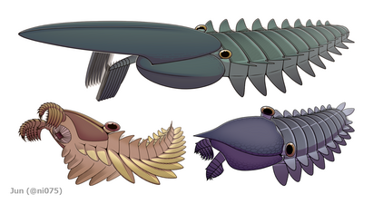
Aegirocassis, Peytoia and Hurdia. Three hurdiid genera which bore dorsal flaps and enlarged head carapace complex.
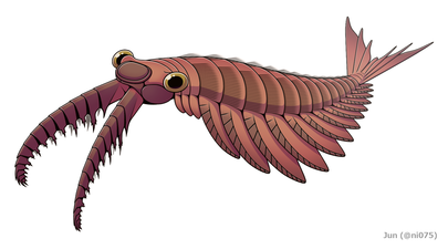
Anomalocaris canadensis, an Anomalocaridid radiodont with small ovoid head carapace complex, 3 pairs of tail fan, and a terminal tailpiece.
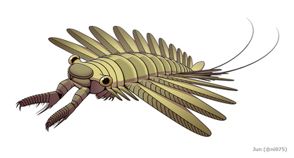
Amplectobelua symbrachiata. An Amplectobeluid radiodont with gnathobase-like structures and two furcae.


Internal structures[]

Traces of muscles, digestive system and nervous system were described from some radiodont fossils. Pairs of well-developed muscles were connected to the ventral flaps located at the lateral cavities of each body segment.[21][9] Between the lateral muscles is a sophisticated digestive system, formed by a widening of the foregut and hindgut, both connected by a narrow midgut associated with 6 pairs of gut divercula (digestive glands).[21][5][23] Compared to the three-segmented brains of euarthropods and two-segmented brains of onychophorans, the brain of radiodonts is composed of only one brain segment originating from the ocular somite, the protocerebrum. The nerves of the frontal appendages and compound eyes arose from the anterior and lateral regions of the brain.[9][15] Posterior to the brain was a pair of apparently unfused ventral nerve cords which ran through the animal's neck region.[9]
Paleoecology[]
Physiology[]
Radiodonts were interpreted as nektonic or animals, with their morphology suggesting an active swimming lifestyle. The muscular, overlapping ventral flaps may have propelled the animal through the water, possibly by moving in a wave-like formation resembling modern rays and cuttlefish.[24] Pairs of dorsal flaps, which make up a tail fan in some species, may have helped steering and/or stabilizing the animal during locomotion.[10][25] In Anomalocaris, morphology of the tail fan even suggests it could rapidly change its swimming direction efficiently.[26] Bands of setal blades with wrinkling lanceolate blades may have increased the surface area, suggesting they were gills, providing the animal's respiratory function.[21][10] Abundance of the remains of scleritzed structures such as disarticulated frontal appendages and head carapace complexes, suggest that mass moulting events may have occurred among radiodonts,[10][3] a behavior which also has been reported in some other Cambrian arthropods such as trilobites.[27]
Diet[]

Radiodonts had diverse feeding strategies, which could be categorized as raptorial predators, sediment sifters, or suspension, filter feeders.[2][28] For example, raptorial predators like Anomalocaris and Amplectobeluids might have been able to catch agile prey by using their raptorial frontal appendages, the latter even bore a robust endite for holding prey like a pincer.[20][17][4] With the smaller head carapace complex and large surface of arthrodial membranes, frontal appendages of these taxa had greater flexibility.[11] Stout frontal appendages of sediment sifters like Hurdia and Peytoia have serrated endites with mesial curvature, which could form a basket-like trap for raking through sediment and passing food items towards the well-developed oral cone.[3] Some species may have other features significantly specialized for a sediment sifting lifestyle, such as Cambroraster with its dome-like H-element similar to the carapace of a horseshoe crab.[3] Endites of frontal appendages from suspension/filter feeders like Tamisiocaris and Aegirocassis have flexible, densely-packed auxiliary spines, which could filter out organic components such as mesozooplankton and phytoplankton down to 0.5mm.[7][10] Frontal appendages of Caryosyntrips, which are unusual for radiodonts in having the direction of endite-bearing surfaces opposing one another and may have been able to manipulate and crush prey in a scissor-like slicing or grasping motion.[17][29]

Peytoia nathorsti, a sediment-sifting hurdiid radiodont with stout frontal appendages, closely associated with a tetraradial oral cone.

Frontal appendage of the suspension-feeding radiodont Tamisiocaris borealis, showing elongated endite with densely-packed auxiliary spines.
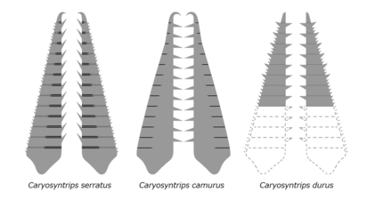
Paired frontal appendages of Caryosyntrips, showing opposed direction.


Oral cones of radiodonts may have been used for suction and/or biting.[18][28][3] Together with the great variety of frontal appendages in different species of radiodonts, differentiation of oral cones between species suggests preferences of different diets as well.[28] For example, the triradial oral cone of Anomalocaris with irregular, tuberculated toothplates and a small opening may have been adapted to small and nektonic prey, [18]; while the rigid tetraradial oral cones of Peytoia, Hurdia and one isolated oral cone attributed to Cambroraster with a larger opening and sometimes additional tooth plates may have been capable to consume larger food items relative to their body size and probably benthic or endobenthic prey. [18] [28][3]
Classification[]
Taxomomic affinities[]
| ||||||||||||||||||||||||||||||||||||||||||||||||
| Summarized phylogeny between Radiodonta and other Ecdysozoan taxa.[30] |


Most analyses suggest that radiodonts are stem-group arthropods[8][7][9][10][2][3] and sister to , a clade including upper stem (e.g. fuxianhuiids and bivalved arthropods) and crown Euarthropoda (e.g. Artiopoda, Chelicerata and Mandibulata).[30] This interpretation is supported by numerous arthropod-like features found on radiodonts, such as compound eyes,[19] digestive glands,[23] arthropodization (on frontal appendages),[30][3] trunk appendages forming by dorsal and ventral elements (precursor of arthropod biramus appendages),[10][3] cuticularized foregut and hindgut,[21] as well as protocerebral anterior sclerite.[31] The constricted neck region with feeding appendicular structures of some radiodont may also shed light on the origin of the sophisticated arthropod head, which is formed by the fusion of multiple anterior body segments.[4][15]

The 'gilled lobopodians' Opabinia (top), Pambdelurion (bottom left) and Kerygmachela (bottom right).

The siberiid lobopodians Siberion (upper left), Megadictyon (bottom center) and Jianshanopodia (upper right).


Taxa just basal to the radiodont and euarthropod branch are Pambdelurion, Kerygmachela and Opabinia, three radiodont-like dinocaridid genera usually referred as 'gilled lobopodians'.[30][10] They have body flaps, digestive glands and specialized frontal appendages like radiodont, but the frontal appendages are not arthropodized and they bore lobopod underneath each of their flaps.[10] Opabinia is more derived by having radiodont-like stalked eyes, tail fan, setal blades, and even euarthropod-like posterior mouth opening.[30] Taxa even basal to 'gilled lobopodians' are siberiids like Megadictyon and Jianshanopodia,[30] a group of lobopodians bore robust frontal appendages and digestive glands, but no body flaps. Such intermediate forms between lobopodian and radiodont/euarthropod suggest that the total-group arthropoda arose from a paraphyletic lobopodian grade, alongside the other two extant panarthropod phyla Tardigrada and Onychophora.[15]


Previous studies may suggest radiodonts as a group other than stem-arthropods, such as cycloneuralian worms undergone convergent with arthropods (based on the cycloneuralian-like radial mouthparts);[32] stem chelicerates alongside megacheirans a.k.a great appendage arthropods (based on the similarity between radiodont frontal appendages, megacheiran great appendages and chelicerae);[33] or Schinderhannes bartelsi, which resolved as a hurdiid radiodont in recent analyses,[30][7][10][2][3] as a species more closely related to euarthropods than other radiodonts (based on some putative euarthropod-like features found on Schinderhannes).[25] However, neither each of them were supported by later investigations. The radial mouthparts are not cycloneuralian-exclusive and more likely present result of convergent evolution or ecdysozoan plesimorphy, since they also have been found in panarthropods such as tardigrade and some lobopodians;[34] the megacheiran great appendages were considered to be deutocerebral,[35][36] which are non-homologous to the radiodont protocerebral frontal appendages;[9][15] putative euarthropod characters found on the single Schinderhannes fossil is questionable and may present other radiodont-like structures.[30]
Interrelationships[]
Traditionally, all radiodont species have been placed within one family, Anomalocarididae,[6] hence the common name 'anomalocaridid'[20][8] and it was still occasionally used to refer the whole order even after reclassification.[9][10] Since the reassignment done by Vinther et al. 2014, most of the radiodont species were reclassified within three new families: Amplectobeluidae, Tamisiocarididae[2][3] (formerly Cetiocaridae[7]), and Hurdiidae.[7][10][2][3] Including Anomalocarididae, the four recent radiodont families may form the clade Anomalocarida.[7]

The original description of the order Radiodonta included Anomalocaris, Laggania (later known as Peytoia), Hurdia, Proboscicaris, Amplectobelua, Cucumericrus, and Parapeytoia.[6] However, Proboscicaris is now regarded as a junior synonym of Hurdia, and Parapeytoia is considered to be a megacheiran.[8][21][10] The position of Cucumericrus within Radiodonta is unclear, as it was either unselected by phylogenetic analysis[3] or resolved in a polytomy with Radiodonta and Euarthropoda.[10]


Under Radiodonta, Caryosyntrips is the basal-most genus alongside Cucumericrus (if included). The genus Anomalocaris always found to be non-monophyletic, usually with Anomalocaris kunmingensis and Anomalocaris briggsi resolved as a member of Amplectobeluidae and Tamisiocarididae respectively.[7][9][10][2][3] Interrelationship of Amplectobeluidae is uncertain, as the amplectobeluid affinities of Lyrarapax and Ramskoeldia were occasionally questioned.[1][3] Monophyly of the speciose family Hurdiidae is well-supported by several derived characters (e.g. distal articulated region of frontal appendage with proximal 5 podomeres bearing subequal endites[16][3]), with Tamisiocarididae often suggested to be its sister group.[7][10][2][3]
- Radiodonta
- ?Cucumericrus
- Caryosyntrips
- Anomalocarida
- Paranomalocaris (placed within Anomalocarididae by some studies.[11])
- (placed within Amplectobeluidae by some studies.[2])
- Anomalocarididae
- Anomalocaris (some species may placed within the other families.[7][10])
- Amplectobeluidae
- Lyrarapax (position questioned by some studies.[1])
- Amplectobelua
- Ramskoeldia (position questioned by some studies.[3])
- Tamisiocarididae
- Hurdiidae
- Aegirocassis
- Peytoia
- Schinderhannes
- Hurdia
- Ursulinacaris
- Stanleycaris
- Cambroraster
- ?
- [37]
- [38]
The first in-depth phylogenetic analysis of Radiodonta was conducted by Vinther et al. in 2014, and it was expanded by Cong et al. later that year by the addition of Lyrarapax unguispinus.[9] The analysis was further modified in 2015 by Van Roy et al. with modified characters and the inclusion of Cucumericrus decoratus and Aegirocassis benmoulai.[10]
|


| Species | Describers | Year Named | Family | Age | Location | Frontal Appendage |
|---|---|---|---|---|---|---|
| Cucumericrus decoratus | Hou, Bergström, & Ahlberg | 1995 | Unknown | |||
| Caryosyntrips serratus | Daley & Budd | 2010 | Wuliuan–Drumian | 
| ||
| Caryosyntrips camurus | Pates & Daley | 2017 | Wuliuan | 
| ||
| Caryosyntrips durus | Pates & Daley | 2017 | Drumian | 
| ||
| Paranomalocaris multisegmentalis | Wang, Huang, & Hu | 2013 | 
| |||
| Paranomalocaris simplex | Jiao, Pates, Lerosey-Aubril, Ortega-Hernandez, Yang, Lan, Zhang | 2021 | 
| |||
| Laminacaris chimera | Guo, Pates, Cong, Daley, Edgecombe, Chen, & Hou | 2018 | 
| |||
| Anomalocaris canadensis | Whiteaves | 1892 | Anomalocarididae | 
| ||
| Lenisicaris pennsylvanica | Resser | 1929 | Anomalocarididae | Cambrian Stage 3 | 
| |
| Lenisicaris lupata | Wu, Ma, Lin, Sun, Zhang, & Fu | 2021 | Anomalocarididae | Cambrian Stage 4 | 
| |
| Anomalocaris kunmingensis | Wang, Huang, & Hu | 2013 | 
| |||
| Houcaris magnabasis | Pates, Daley, Edgecombe, Cong & Lieberman | 2019 | Tamisiocarididae | Cambrian Stage 4 | 
| |
| Houcaris saron | Hou, Bergström, & Ahlberg | 1995 | Tamisiocarididae | Cambrian Stage 3 | 
| |
| Anomalocaris briggsi | Nedin | 1995 | Tamisiocarididae | 
| ||
| Ramskoeldia platyacantha | Cong, Edgecombe, Daley, Guo, Pates, & Hou | 2018 | Amplectobeluidae | 
| ||
| Ramskoeldia consimilis | Cong, Edgecombe, Daley, Guo, Pates, & Hou | 2018 | Amplectobeluidae | 
| ||
| Lyrarapax unguispinus | Cong, Ma, Hou, Edgecombe, & Strausfield | 2014 | Amplectobeluidae | Cambrian Stage 3 | 
| |
| Lyrarapax trilobus | Cong, Daley, Edgecombe, Hou, & Chen | 2016 | Amplectobeluidae | Cambrian Stage 3 | 
| |
| Amplectobelua symbrachiata | Hou, Bergström, & Ahlberg | 1995 | Amplectobeluidae | 
| ||
| Amplectobelua stephenensis | Daley & Budd | 2010 | Amplectobeluidae | 
| ||
| Tamisiocaris borealis | Daley & Peel | 2010 | Tamisiocarididae | 
| ||
| Ursulinacaris grallae | Pates, Daley & Butterfield | 2019 | Hurdiidae | Wuliuan | 
| |
| Schinderhannes bartelsi | Kühl, Briggs, & Rust | 2009 | Hurdiidae | Emsian | ||
| Stanleycaris hirpex | Pates, Daley, & Ortega-Hernández | 2018 | Hurdiidae | 
| ||
| Peytoia nathorsti | Walcott | 1911 | Hurdiidae | 
| ||
| Peytoia infercambriensis | Lendzion | 1975 | Hurdiidae | Cambrian Stage 3 | 
| |
| Aegirocassis benmoulai | Van Roy, Daley, & Briggs | 2015 | Hurdiidae | Tremadocian | 
| |
| Hurdia victoria | Walcott | 1912 | Hurdiidae | Wuliuan–Drumian | 
| |
| Hurdia triangulata | Walcott | 1912 | Hurdiidae | Wuliuan |
| |
| Cambroraster falcatus | Moysiuk & Caron | 2019 | Hurdiidae | Wuliuan | 
| |
| Pahvantia hastata | Robison & Richards | 1981 | Hurdiidae | Drumian | 
| |
| Cordaticaris striatus | Sun, Zeng, & Zhao | 2020 | Hurdiidae | Drumian | ||
| Zhenghecaris shankouensis | Vanner, Chen, Huang, Charbonnier, & Wang | 2006 | Hurdiidae | Unknown | ||
| Buccaspinea cooperi | Pates, Lerosey-Aubril, Daley, Kier, Bonino & Ortega-Hernández | 2021 | Hurdiidae | Drumian | 
| |
| Titanokorys gainesi | Caron & Moysiuk | 2021 | Hurdiidae | Wuliuan | 
|
History[]
The history of radiodonts is complex. Incomplete specimens pertaining to different body parts of the same species had historically been interpreted as belonging to different species and even different phyla.[6][8] Prior to their recognition as a group, radiodont specimens had been assigned to five different phyla: Porifera, Cnidaria, Echinodermata, Annelida, and Arthropoda.[6]
The first known radiodont specimens were collected from the trilobite beds of Mount Stephen by Richard G. McConnell of the Geological Survey of Canada in 1886[6] or 1888.[39] These specimens were named Anomalocaris canadensis in 1892 by GSC paleontologist Joseph Whiteaves.[39] Whiteaves interpreted the specimens, now known to be isolated frontal appendages, as the abdomen of a phyllocarid crustacean. Additional radiodont specimens were described in 1911 by Charles Walcott. He interpreted an isolated oral cone, which he named Peytoia, as a jellyfish, and a poorly-preserved but relatively complete specimen, which he named Laggania, as a holothurian. In 1912 Walcott named Hurdia based on an isolated h-element, which he interpreted as the carapace of a crustacean. A Hurdia p-element was named Proboscicaris in 1962, and interpreted as the carapace of a bivalved arthropod.
The Geological Survey of Canada initiated a revision of Burgess Shale fossils in 1966, overseen by Cambridge University paleontologist Harry B. Whittington.[6] This revision would ultimately lead to the discovery of the complete radiodont body plan. In 1978, Simon Conway Morris recognized that the mouthparts of Laggania were Peytoia-like, but he interpreted this as evidence that it was a composite fossil made up of a Peytoia jellyfish and a sponge.[40] In 1979, Derek Briggs recognized that the fossils of Anomalocaris were appendages, not abdomens, but interpreted them as walking legs.[41] It was not until 1985 that the true nature of the fossils of Anomalocaris, Laggania, and Peytoia was recognized, and they were all assigned to a single genus, Anomalocaris. Subsequently, it was recognized that Anomalocaris was a distinct form from the other two, resulting in a split into two genera, the latter of which was variously named Laggania and Peytoia until it was determined that Peytoia had priority. It was later recognized that some of the fossils assigned to these taxa belonged to another form, which was recognized as bearing a carapace made up of Hurdia and Proboscicaris elements. Finally, in 2009, these specimens were redescribed as Hurdia.[8]
The taxon Radiodonta itself was coined in 1996 by Desmond Collins, after it was established that Anomalocaris and its kin represented a distinctive lineage with arthropod affinities rather than a hitherto unknown phylum.[6] Collins also established the class Dinocarida to contain the order Radiodonta as well as the Opabiniidae, which he recognized as distinct due to its lacking the distinctive oral cone structure of radiodonts. Radiodonta was first given a phylogenetic definition in 2014.[7] Radiodonta was originally viewed as containing a single family, Anomalocarididae, but it was divided into four families in 2014: Amplectobeluidae, Anomalocarididae, Cetiocaridae, and Hurdiidae.[7] The name Cetiocaridae did not conform to the International Code of Zoological Nomenclature and so was renamed Tamisiocarididae in 2019.[42]
Until the 2010s, radiodonts were typically considered to be uniformly large apex predators, but discoveries of new species over the course of that decade led to a considerable increase in the known ecological and morphological diversity of the group.[43]
References[]
- ^ Jump up to: a b c d e f Cong, Pei-Yun; Edgecombe, Gregory D.; Daley, Allison C.; Guo, Jin; Pates, Stephen; Hou, Xian-Guang (2018). "New radiodonts with gnathobase-like structures from the Cambrian Chengjiang biota and implications for the systematics of Radiodonta". Papers in Palaeontology. 4 (4): 605–621. doi:10.1002/spp2.1219. ISSN 2056-2802.
- ^ Jump up to: a b c d e f g h i j k l m Lerosey-Aubril, Rudy; Pates, Stephen (2018-09-14). "New suspension-feeding radiodont suggests evolution of microplanktivory in Cambrian macronekton". Nature Communications. 9 (1): 3774. doi:10.1038/s41467-018-06229-7. ISSN 2041-1723. PMC 6138677. PMID 30218075.
- ^ Jump up to: a b c d e f g h i j k l m n o p q r s t u v w x y z aa ab ac ad ae af ag Moysiuk, J.; Caron, J.-B. (2019-08-14). "A new hurdiid radiodont from the Burgess Shale evinces the exploitation of Cambrian infaunal food sources". Proceedings of the Royal Society B: Biological Sciences. 286 (1908): 20191079. doi:10.1098/rspb.2019.1079. PMC 6710600. PMID 31362637.
- ^ Jump up to: a b c d e f g Cong, Peiyun; Daley, Allison C.; Edgecombe, Gregory D.; Hou, Xianguang (2017-08-30). "The functional head of the Cambrian radiodontan (stem-group Euarthropoda) Amplectobelua symbrachiata". BMC Evolutionary Biology. 17 (1): 208. doi:10.1186/s12862-017-1049-1. ISSN 1471-2148. PMC 5577670. PMID 28854872.
- ^ Jump up to: a b c d Cong, Peiyun; Daley, Allison C.; Edgecombe, Gregory D.; Hou, Xianguang; Chen, Ailin (September 2016). "Morphology of the radiodontan Lyrarapax from the early Cambrian Chengjiang biota". Journal of Paleontology. 90 (4): 663–671. doi:10.1017/jpa.2016.67. ISSN 0022-3360. S2CID 88742430.
- ^ Jump up to: a b c d e f g h i j k Collins, Desmond (1996). "The "evolution" of Anomalocaris and its classification in the arthropod class Dinocarida (nov.) and order Radiodonta (nov.)". Journal of Paleontology. 70 (2): 280–293. doi:10.1017/S0022336000023362.
- ^ Jump up to: a b c d e f g h i j k l m n Vinther, Jakob; Stein, Martin; Longrich, Nicholas R.; Harper, David A. T. (2014). "A suspension-feeding anomalocarid from the Early Cambrian" (PDF). Nature. 507 (7493): 496–499. doi:10.1038/nature13010. PMID 24670770. S2CID 205237459.
- ^ Jump up to: a b c d e f g h i Daley, Allison C.; Budd, Graham E.; Caron, Jean-Bernard; Edgecombe, Gregory D.; Collins, Desmond (2009). "The Burgess Shale anomalocaridid Hurdia and its significance for early euarthropod evolution". Science. 323 (5921): 1597–1600. doi:10.1126/science.1169514. PMID 19299617. S2CID 206517995.
- ^ Jump up to: a b c d e f g h i j Cong, Peiyun; Ma, Xiaoya; Hou, Xianguang; Edgecombe, Gregory D.; Strausfeld, Nicholas J. (2014). "Brain structure resolves the segmental affinity of anomalocaridid appendages". Nature. 513 (7519): 538–42. doi:10.1038/nature13486. PMID 25043032. S2CID 4451239.
- ^ Jump up to: a b c d e f g h i j k l m n o p q r s t u v w x y z aa Van Roy, Peter; Daley, Allison C.; Briggs, Derek E. G. (2015). "Anomalocaridid trunk limb homology revealed by a giant filter-feeder with paired flaps". Nature. 522 (7554): 77–80. doi:10.1038/nature14256. PMID 25762145. S2CID 205242881.
- ^ Jump up to: a b c d e Liu, Jianni; Lerosey-Aubril, Rudy; Steiner, Michael; Dunlop, Jason A.; Shu, Degan; Paterson, John R. (2018-11-01). "Origin of raptorial feeding in juvenile euarthropods revealed by a Cambrian radiodontan". National Science Review. 5 (6): 863–869. doi:10.1093/nsr/nwy057. ISSN 2095-5138.
- ^ Pates, Stephen; Botting, Joseph P.; McCobb, Lucy M. E.; Muir, Lucy A. (2020). "A miniature Ordovician hurdiid from Wales demonstrates the adaptability of Radiodonta". Royal Society Open Science. 7 (6): 200459. doi:10.1098/rsos.200459. ISSN 2054-5703.
- ^ Wu, Yu; Fu, Dongjing; Ma, Jiaxin; Lin, Weiliang; Sun, Ao; Zhang, Xingliang (2021-06-01). "Houcaris gen. nov. from the early Cambrian (Stage 3) Chengjiang Lagerstätte expanded the palaeogeographical distribution of tamisiocaridids (Panarthropoda: Radiodonta)". PalZ. 95 (2): 209–221. doi:10.1007/s12542-020-00545-4. ISSN 1867-6812.
- ^ Caron, J.-B.; Moysiuk, J. (September 2021). "A giant nektobenthic radiodont from the Burgess Shale and the significance of hurdiid carapace diversity". Royal Society Open Science. 8 (9): 210664. doi:10.1098/rsos.210664.
- ^ Jump up to: a b c d e f g Ortega-Hernández, Javier; Janssen, Ralf; Budd, Graham E. (2017-05-01). "Origin and evolution of the panarthropod head – A palaeobiological and developmental perspective". Arthropod Structure & Development. Evolution of Segmentation. 46 (3): 354–379. doi:10.1016/j.asd.2016.10.011. ISSN 1467-8039. PMID 27989966.
- ^ Jump up to: a b c d e Pates, Stephen; Daley, Allison C.; Butterfield, Nicholas J. (2019-06-11). "First report of paired ventral endites in a hurdiid radiodont". Zoological Letters. 5 (1): 18. doi:10.1186/s40851-019-0132-4. ISSN 2056-306X. PMC 6560863. PMID 31210962.
- ^ Jump up to: a b c d Daley, Allison C.; Budd, Graham E. (2010). "New anomalocaridid appendages from the Burgess Shale, Canada". Palaeontology. 53 (4): 721–738. doi:10.1111/j.1475-4983.2010.00955.x. ISSN 1475-4983.
- ^ Jump up to: a b c d Daley, Allison C.; Bergström, Jan (April 2012). "The oral cone of Anomalocaris is not a classic peytoia". Naturwissenschaften. 99 (6): 501–504. doi:10.1007/s00114-012-0910-8. ISSN 0028-1042. PMID 22476406. S2CID 2042726.
- ^ Jump up to: a b Strausfeld, Nicholas J.; Ma, Xiaoya; Edgecombe, Gregory D.; Fortey, Richard A.; Land, Michael F.; Liu, Yu; Cong, Peiyun; Hou, Xianguang (August 2015). "Arthropod eyes: The early Cambrian fossil record and divergent evolution of visual systems". Arthropod Structure & Development. 45 (2): 152–172. doi:10.1016/j.asd.2015.07.005. PMID 26276096.
- ^ Jump up to: a b c d Xian‐Guang, Hou; Bergström, Jan; Ahlberg, Per (September 1995). "Anomalocaris and other large animals in the lower Cambrian Chengjiang fauna of southwest China". GFF. 117 (3): 163–183. doi:10.1080/11035899509546213. ISSN 1103-5897.
- ^ Jump up to: a b c d e f g h Daley, Allison C.; Edgecombe, Gregory D. (2014). "Morphology of Anomalocaris canadensis from the Burgess Shale". Journal of Paleontology. 88 (1): 68–91. doi:10.1666/13-067. S2CID 86683798.
- ^ Van Roy, Peter; Daley, Allison C.; Briggs, Derek E. G. (2013). Anomalocaridids had two sets of lateral flaps. 57th Annual Meeting of The Paleontological Association. Zurich, Switzerland.
- ^ Jump up to: a b Vannier, Jean; Liu, Jianni; Lerosey-Aubril, Rudy; Vinther, Jakob; Daley, Allison C. (2014-05-02). "Sophisticated digestive systems in early arthropods". Nature Communications. 5 (1): 3641. doi:10.1038/ncomms4641. ISSN 2041-1723. PMID 24785191.
- ^ Usami, Yoshiyuki (2006-01-07). "Theoretical study on the body form and swimming pattern of Anomalocaris based on hydrodynamic simulation". Journal of Theoretical Biology. 238 (1): 11–17. doi:10.1016/j.jtbi.2005.05.008. ISSN 0022-5193. PMID 16002096.
- ^ Jump up to: a b Kühl, Gabriele; Briggs, Derek E. G.; Rust, Jes (2009-02-06). "A Great-Appendage Arthropod with a Radial Mouth from the Lower Devonian Hunsrück Slate, Germany". Science. 323 (5915): 771–773. doi:10.1126/science.1166586. ISSN 0036-8075. PMID 19197061. S2CID 47555807.
- ^ Sheppard, K. A.; Rival, D. E.; Caron, J.-B. (2018-10-01). "On the Hydrodynamics of Anomalocaris Tail Fins". Integrative and Comparative Biology. 58 (4): 703–711. doi:10.1093/icb/icy014. ISSN 1540-7063. PMID 29697774.
- ^ Daley, Allison; Drage, Harriet (2015-09-01). "The fossil record of ecdysis, and trends in the moulting behaviour of trilobites". Arthropod Structure & Development. 45 (2): 71–96. doi:10.1016/j.asd.2015.09.004. PMID 26431634.
- ^ Jump up to: a b c d De Vivo, Giacinto; Lautenschlager, Stephan; Vinther, Jakob (2016-12-16). "Reconstructing anomalocaridid feeding appendage dexterity sheds light on radiodontan ecology". Cite journal requires
|journal=(help) - ^ Pates, S.; Daley, A. C. (2017). "Caryosyntrips: a radiodontan from the Cambrian of Spain, USA and Canada". Papers in Palaeontology. 3 (3): 461–470. doi:10.1002/spp2.1084. ISSN 2056-2802.
- ^ Jump up to: a b c d e f g h i Ortega-Hernández, Javier (Dec 2014). "Making sense of 'lower' and 'upper' stem-group Euarthropoda, with comments on the strict use of the name Arthropoda von Siebold, 1848". Biological Reviews of the Cambridge Philosophical Society. 91 (1): 255–273. doi:10.1111/brv.12168. ISSN 1469-185X. PMID 25528950. S2CID 7751936.
- ^ Ortega-Hernández, Javier (2015-06-15). "Homology of Head Sclerites in Burgess Shale Euarthropods". Current Biology. 25 (12): 1625–1631. doi:10.1016/j.cub.2015.04.034. ISSN 0960-9822. PMID 25959966.
- ^ Xian‐Guang, Hou; Bergström, Jan; Ahlberg, Per (September 1995). "Anomalocaris and other large animals in the lower Cambrian Chengjiang fauna of southwest China". GFF. 117 (3): 163–183. doi:10.1080/11035899509546213. ISSN 1103-5897.
- ^ Haug, Joachim T.; Waloszek, Dieter; Maas, Andreas; Liu, Yu; Haug, Carolin (March 2012). "Functional morphology, ontogeny and evolution of mantis shrimp-like predators in the Cambrian: MANTIS SHRIMP-LIKE CAMBRIAN PREDATORS". Palaeontology. 55 (2): 369–399. doi:10.1111/j.1475-4983.2011.01124.x.
- ^ Smith, Martin R.; Caron, Jean-Bernard (June 2015). "Hallucigenia 's head and the pharyngeal armature of early ecdysozoans" (PDF). Nature. 523 (7558): 75–78. doi:10.1038/nature14573. ISSN 1476-4687. PMID 26106857. S2CID 205244325.
- ^ Tanaka, Gengo; Hou, Xianguang; Ma, Xiaoya; Edgecombe, Gregory D.; Strausfeld, Nicholas J. (October 2013). "Chelicerate neural ground pattern in a Cambrian great appendage arthropod". Nature. 502 (7471): 364–367. doi:10.1038/nature12520. ISSN 1476-4687. PMID 24132294. S2CID 4456458.
- ^ Ortega-Hernández, Javier; Lerosey-Aubril, Rudy; Pates, Stephen (2019-12-18). "Proclivity of nervous system preservation in Cambrian Burgess Shale-type deposits". Proceedings of the Royal Society B: Biological Sciences. 286 (1917): 20192370. doi:10.1098/rspb.2019.2370. PMC 6939931. PMID 31822253.
- ^ Sun, Zhixin; Zeng, Han; Zhao, Fangchen (2020-08-01). "A new middle Cambrian radiodont from North China: Implications for morphological disparity and spatial distribution of hurdiids". Palaeogeography, Palaeoclimatology, Palaeoecology. 558: 109947. doi:10.1016/j.palaeo.2020.109947. ISSN 0031-0182.
- ^ Pates, Stephen; Lerosey-Aubril, Rudy; Daley, Allison C.; Kier, Carlo; Bonino, Enrico; Ortega-Hernández, Javier (2021-01-19). "The diverse radiodont fauna from the Marjum Formation of Utah, USA (Cambrian: Drumian)". PeerJ. 9: e10509. doi:10.7717/peerj.10509. ISSN 2167-8359.
- ^ Jump up to: a b Whiteaves, J. F. (1892). "Description of a new genus and species of phyllocarid Crustacea from the Middle Cambrian of Mount Stephen, B. C.". The Canadian Record of Science. 5 (4).
- ^ Conway Morris, S. (1978). "Laggania cambria Walcott: A Composite Fossil". Journal of Paleontology. 52 (1): 126–131. JSTOR 1303799.
- ^ Briggs, D. E. G. (1979). "Anomalocaris, the largest known Cambrian arthropod". Palaeontology. 22 (3): 631–664.
- ^ Pates, Stephen; Daley, Allison C. (2019). "The Kinzers Formation (Pennsylvania, USA): the most diverse assemblage of Cambrian Stage 4 radiodonts". Geological Magazine. 156 (07): 1233–1246. doi:10.1017/S0016756818000547.
- ^ Paterson, John R.; Edgecombe, Gregory D.; García-Bellido, Diego C. (2020). "Disparate compound eyes of Cambrian radiodonts reveal their developmental growth mode and diverse visual ecology". Science Advances. doi:10.1126/sciadv.abc6721. hdl:10141/622906.
| hide | |||||||||||
|---|---|---|---|---|---|---|---|---|---|---|---|
| |||||||||||
| Basal Radiodonts |  | ||||||||||
| Anomalocarida |
| ||||||||||
- Dinocarida
- Prehistoric arthropod orders
- Cambrian Series 2 first appearances
- Early Devonian extinctions