Siphoviridae
| Siphoviridae | |
|---|---|

| |
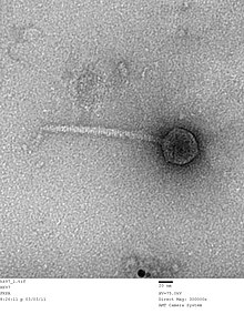
| TME picture of an Escherichia virus HK97 virion, Siphoviridae | |
| Virus classification | |
| (unranked): | Virus |
| Realm: | Duplodnaviria |
| Kingdom: | Heunggongvirae |
| Phylum: | Uroviricota |
| Class: | Caudoviricetes |
| Order: | Caudovirales |
| Family: | Siphoviridae |
| Genera | |
| Synonyms[1] | |
| |
Siphoviridae is a family of double-stranded DNA viruses in the order Caudovirales. Bacteria and archaea serve as natural hosts. There are 1,166 species in this family, assigned to 366 genera and 22 subfamilies.[2][3] The characteristic structural features of this family are a nonenveloped head and noncontractile tail.
Structure[]

Viruses in Siphoviridae are non-enveloped, with icosahedral and head-tail geometries[2] (morphotype B1) or a prolate capsid (morphotype B2), and T=7 symmetry. The diameter is around 60 nm.[2] Members of this family are also characterized by their filamentous, cross-banded, noncontractile tails, usually with short terminal and subterminal fibers. Genomes are double stranded and linear, around 50 kb in length,[2] containing about 70 genes. The guanine/cytosine content is usually around 52%.
Life cycle[]
Viral replication is cytoplasmic. Entry into the host cell is achieved by adsorption into the host cell. Replication follows the replicative transposition model. DNA-templated transcription is the method of transcription. Translation takes place by -1 ribosomal frameshifting, and +1 ribosomal frameshifting. The virus exits the host cell by lysis, and holin/endolysin/spanin proteins.[2] Bacteria and archaea serve as the natural host. Transmission routes are passive diffusion.[2]
Taxonomy[]

The following subfamilies are recognized:[3]
The following genera are unassigned to a subfamily:[3]
References[]
- ^ Safferman, R.S.; Cannon, R.E.; Desjardins, P.R.; Gromov, B.V.; Haselkorn, R.; Sherman, L.A.; Shilo, M. (1983). "Classification and Nomenclature of Viruses of Cyanobacteria". Intervirology. 19 (2): 61–66. doi:10.1159/000149339. PMID 6408019.
- ^ a b c d e f "Viral Zone". ExPASy. Retrieved 1 July 2015.
- ^ a b c "Virus Taxonomy: 2020 Release". International Committee on Taxonomy of Viruses (ICTV). March 2021. Retrieved 12 May 2021.
- ^ Lood R, Mörgelin M, Holmberg A, Rasmussen M, Collin M (2008). "Inducible Siphoviruses in superficial and deep tissue isolates of Propionibacterium acnes". BMC Microbiol. 8: 139. doi:10.1186/1471-2180-8-139. PMC 2533672. PMID 18702830.
External links[]
| Wikispecies has information related to Siphoviridae. |
- Siphoviridae
- Virus families