Fascial compartments of leg
| Fascial compartments of leg | |
|---|---|
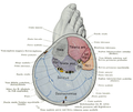
 Cross-section through middle of leg. Four compartments painted in different colors. | |
 Muscles of the lower leg. Four compartments painted in different colors. | |
| Anatomical terminology |
The fascial compartments of the leg are the four fascial compartments that separate and contain the muscles of the lower leg (from the knee to the ankle). The compartments are divided by septa formed from the fascia. The compartments usually have nerve and blood supplies separate from their neighbours. All of the muscles within a compartment will generally be supplied by the same nerve.
Intermuscular septa[]
The lower leg is divided into four compartments by the interosseous membrane of the leg, the anterior intermuscular septum, the transverse intermuscular septum and the posterior intermuscular septum.[1]
Each compartment contains connective tissue, nerves and blood vessels. The septa are formed from the fascia which is made up of a strong type of connective tissue. The fascia also separates the skeletal muscles from the subcutaneous tissue.[2] Due to the great pressure placed on the leg, from the column of blood from the heart to the feet, the fascia is very thick in order to support the leg muscles.[3] The thickness of the fascia can give problems when any inflammation present in the leg has little room to expand into. Blood vessels and nerves can also be affected by the pressure caused by any swelling in the leg. If the pressure becomes great enough, blood flow to the muscle can be blocked, leading to a condition known as compartment syndrome. Severe damage to the nerve and blood vessels around a muscle can cause the muscle to die and amputation might be necessary.[4]
Compartments[]
| Image | Compartment | Muscles | Neurovascular structures |
|---|---|---|---|

|
Anterior compartment |
|
Deep fibular (peroneal) nerve and anterior tibial vessels |

|
Lateral compartment | Superficial fibular (peroneal) nerve and fibular artery | |

|
Deep posterior compartment |
|
Tibial nerve, posterior tibial artery and posterior tibial vessels such as the fibular artery |

|
Superficial posterior compartment |
|
Tibial nerve |
Additional images[]

Animation
Cross-section of the right leg.


See also[]
- Fascial compartments of thigh
- Fascial compartments of arm
- Fascial compartments of forearm
References[]
- ^ Fraipont, Michael J.; Adamson, Gregory J. (2003). "Chronic Exertional Compartment Syndrome". The Journal of the American Academy of Orthopaedic Surgeons. 11 (4): 268–76. doi:10.5435/00124635-200307000-00006. PMID 12889865. S2CID 31965215.
- ^ Saladin, Kenneth S. (2012). Anatomy and Physiology: The Unity of Form and Function. New York: McGraw Hill. p. 315.
- ^ http://www.amazonhmt.com/veins_work.html[full citation needed]
- ^ MedlinePlus Encyclopedia: Compartment syndrome
External links[]
| Wikimedia Commons has media related to Fascial compartments of the leg. |
- Muscles of the lower limb