Peroneus tertius
| Peroneus tertius | |
|---|---|
 Animation | |
| Details | |
| Origin | distal anterior surface of the fibula also the interosseous membrane |
| Insertion | dorsal surface of metatarsal 5 |
| Artery | anterior tibial artery |
| Nerve | deep fibular nerve |
| Actions | dorsiflexion and eversion of the foot |
| Identifiers | |
| Latin | Musculus peronaeus tertius, musculus fibularis tertius |
| TA98 | A04.7.02.039 |
| TA2 | 2649 |
| FMA | 22538 |
| Anatomical terms of muscle | |
The peroneus tertius muscle (also known as the fibularis tertius muscle) is a muscle of the lower limb of the human body. It arises from the anterior surface of the fibula and the interosseous membrane, and inserts into the fifth metatarsal bone. Its presence is variable in humans, and it is likely to be an accessory muscle to bipedalism. The muscle is a weak dorsiflexor of the ankle joint, and an evertor of the foot at the ankle joint. It may be involved in ankle injuries, and may rupture.
Structure[]
The peroneus tertius muscle arises from the lower third of the anterior surface of the fibula (anterior compartment of lower leg), the lower part of the interosseous membrane, and an intermuscular septum between it and the peroneus brevis muscle.[1] The septum is sometimes called the intermuscular septum of Otto.
The tendon passes under the superior extensor retinaculum of foot and inferior extensor retinaculum of foot in the same canal as the extensor digitorum longus muscle.[1] It may be mistaken as a fifth tendon of the extensor digitorum longus muscle.[1] It is inserted into the medial part of the posterior surface of the shaft of the fifth metatarsal bone.[1]
Nerve supply[]
The peroneus tertius muscle is supplied by the deep fibular nerve.[2][3] Rarely, it may also be supplied by the common fibular nerve.[2] This is unlike the other peroneal muscles, which are innervated by the superficial fibular nerve, since the peroneus tertius is a member of the anterior compartment.
Variation[]
The peroneus tertius muscle may be absent in humans.[1][4] It may be absent in as few as 5% of people,[4] or as many as 72% depending on the population surveyed.[1] It is rarely found in other primates, which has linked its function to efficient terrestrial bipedalism.[4]
Function[]
The peroneus tertius muscle is a weak dorsiflexor of the ankle joint, and an evertor of the foot at the ankle joint. It is likely to be helpful in bipedal walking, although not essential.[4]
Clinical significance[]
The peroneus tertius muscle may be involved in ankle injuries.[1] It may rupture.[5] This is caused by hyperextension.[6] In horses, this may cause the Achilles tendon to have a slight dip.[7]
The peroneus tertius muscle may be imaged using medical ultrasound.[6]
History[]
The peroneus tertius muscle is also known as the fibularis tertius muscle.[5]
Other animals[]
The peroneus tertius muscle in horses originates from the near the lateral condyle of the femur, passes through the extensor sulcus on the head of the tibia, and inserts onto the third metatarsal bone, the third and fourth tarsal bones, and the calcaneus.[7]
Additional images[]
This gallery of anatomic features needs cleanup to abide by the medical manual of style. |

Muscles of the front of the leg. (peroneus tertius visible at center left)
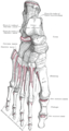
Bones of the right foot (dorsal surface).

The mucous sheaths of the tendons around the ankle (lateral aspect).

Dorsum of Foot. Deep dissection.

Dorsum of Foot. Deep dissection.


See also[]
References[]
![]() This article incorporates text in the public domain from page 482 of the 20th edition of Gray's Anatomy (1918)
This article incorporates text in the public domain from page 482 of the 20th edition of Gray's Anatomy (1918)
- ^ a b c d e f g Salem, Abdel Halim; Abdel Kader, Ghada; Almallah, Amani A.; Hussein, Hoda H.; Abdel Badie, Ahmed; Behbehani, Nadiah; Nedham, Fatema N.; Nedham, Ayesha N.; Almarshad, Reem; Alshammari, Munirah; Amer, Hanine; Hasan, Wafa A.; Alyaseen, Farah A.; Mohammed, Elaf A. (2018-11-01). "Variations of peroneus tertius muscle in five Arab populations: A clinical study". Translational Research in Anatomy. 13: 1–6. doi:10.1016/j.tria.2018.11.001. ISSN 2214-854X.
- ^ a b Zetaruk, Merrilee; Hyman, Jeff (2007-01-01), Frontera, Walter R.; Herring, Stanley A.; Micheli, Lyle J.; Silver, Julie K (eds.), "CHAPTER 32 - Leg Injuries", Clinical Sports Medicine, Edinburgh: W.B. Saunders, pp. 441–457, doi:10.1016/b978-141602443-9.50035-0, ISBN 978-1-4160-2443-9, retrieved 2021-02-23
- ^ Shapiro, L. E.; Kim, J. H.; Lee, S. J.; Yoo, J. J.; Atala, A.; Ko, I. K. (2016). "16 - In Situ Volumetric Muscle Repair". In Situ Tissue Regeneration - Host Cell Recruitment and Biomaterial Design. Academic Press. pp. 295–312. doi:10.1016/B978-0-12-802225-2.00016-7. ISBN 978-0-12-802225-2.
- ^ a b c d Jungers, William L.; Meldrum, D. Jeffrey; Stern, Jack T. (1993-11-01). "The functional and evolutionary significance of the human peroneus tertius muscle". Journal of Human Evolution. 25 (5): 377–386. doi:10.1006/jhev.1993.1056. ISSN 0047-2484.
- ^ a b Guard, Charles L.; Peek, Simon F.; Fecteau, Gilles (2018). "12 - Musculoskeletal Disorders". Rebhun's Diseases of Dairy Cattle (3rd ed.). Saunders. pp. 553–604. doi:10.1016/B978-0-323-39055-2.00012-7. ISBN 978-0-323-39055-2.
- ^ a b Dyson, Sue J. (2011). "80 - Other Soft Tissue Injuries". Diagnosis and Management of Lameness in the Horse (2nd ed.). Saunders. pp. 802–806. doi:10.1016/B978-1-4160-6069-7.00080-8. ISBN 978-1-4160-6069-7.
- ^ a b Walmsley, John P. (2011). "46 - The Stifle". Diagnosis and Management of Lameness in the Horse (2nd ed.). Saunders. pp. 532–549. doi:10.1016/B978-1-4160-6069-7.00046-8. ISBN 978-1-4160-6069-7.
External links[]
| Wikimedia Commons has media related to Peroneus tertius. |
- Anatomy photo:15:st-0411 at the SUNY Downstate Medical Center - "The Leg: Muscles"
- PTCentral
| Authority control: Scientific databases |
|---|
- Wikipedia articles incorporating text from the 20th edition of Gray's Anatomy (1918)
- Muscles of the lower limb