Tibialis posterior muscle
This article includes a list of references, related reading or external links, but its sources remain unclear because it lacks inline citations. (October 2020) |
| Tibialis posterior muscle | |
|---|---|
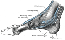 The mucous sheaths of the tendons around the ankle. Medial aspect. (Tibialis posterior labeled at top center.) | |
| Details | |
| Origin | Tibia and fibula |
| Insertion | Navicular and medial cuneiform bone |
| Artery | Posterior tibial artery |
| Nerve | Tibial nerve |
| Actions | Inversion of the foot and plantar flexion of the foot at the ankle |
| Antagonist | Fibularis brevis and longus, antagonist to the inversion. |
| Identifiers | |
| Latin | Musculus tibialis posterior |
| TA98 | A04.7.02.051 |
| TA2 | 2666 |
| FMA | 51099 |
| Anatomical terms of muscle | |
The tibialis posterior muscle is the most central of all the leg muscles, and is located in the deep posterior compartment of the leg. It is the key stabilizing muscle of the lower leg.
Structure[]
The tibialis posterior muscle originates on the inner posterior border of the fibula laterally.[1] It is also attached to the interosseous membrane medially, which attaches to the tibia and fibula.[1]
The tendon of the tibialis posterior muscle (sometimes called the posterior tibial tendon) descends posterior to the medial malleolus.[1] It terminates by dividing into plantar, main, and recurrent components. The main portion inserts into the tuberosity of the navicular bone.[1] The smaller portion inserts into the plantar surface of the medial cuneiform. The plantar portion inserts into the bases of the second, third and fourth metatarsals, the intermediate and lateral cuneiforms and the cuboid. The recurrent portion inserts into the sustentaculum tali of the calcaneus.
Blood is supplied to the muscle by the posterior tibial artery.
Nerve supply[]
The tibialis posterior muscle is suppled by the tibial nerve.
Function[]
The tibialis posterior muscle is a key muscle for stabilization of the lower leg. It also contracts to produce inversion of the foot, and assists in the plantarflexion of the foot at the ankle.[2] The tibialis posterior has a major role in supporting the medial arch of the foot. Dysfunction of the tibialis posterior, including rupture of the tibialis posterior tendon, can lead to flat feet in adults, as well as a valgus deformity due to unopposed eversion when inversion is lost.[3][4]
Clinical significance[]
Injury to the distal tendon of the tibialis posterior muscle is rare.[2] It may be caused during exercise.[2] It usually presents with pain on the medial side of the ankle.[2] This may be treated with dry needling acupuncture.[1]
Additional images[]
This gallery of anatomic features needs cleanup to abide by the medical manual of style. |

Bones of the right leg. Posterior surface.

Bones of the right foot. Plantar surface.

Coronal section through right talocrural and talocalcaneal joints.

Muscles of the back of the leg. Deep layer.

Muscles of the sole of the foot. Third layer.

The popliteal, posterior tibial, and peroneal arteries.

Muscles of the back of the leg. Deep layer.

Muscles of the back of the leg. Deep layer.

Muscles of the leg.Posterior view.

Muscles of the sole of the foot.

Dorsum of Foot. Ankle joint. Deep dissection

Dorsum of Foot. Ankle joint. Deep dissection.
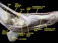
Ankle joint. Deep dissection. Medial view


References[]
- ^ a b c d e Ma, Yun-tao (2011-01-01), Ma, Yun-tao (ed.), "CHAPTER 14 - General Principles of Treating Soft Tissue Dysfunction in Sports Injuries", Acupuncture for Sports and Trauma Rehabilitation, Saint Louis: Churchill Livingstone, pp. 212–233, doi:10.1016/b978-1-4377-0927-8.00014-2, ISBN 978-1-4377-0927-8, retrieved 2021-02-21
- ^ a b c d Hunt, Kenneth J. (2020-01-01), Porter, David A.; Schon, Lew C. (eds.), "10 - Posterior Tibialis Tendon Injury in the Athlete", Baxter's the Foot and Ankle in Sport (Third Edition), Philadelphia: Elsevier, pp. 206–223, doi:10.1016/b978-0-323-54942-4.00010-5, ISBN 978-0-323-54942-4, retrieved 2021-02-21
- ^ Durrant, B., Chockalingam, N. and Hashmi, F., 2011. Posterior tibial tendon dysfunction: a review. Journal of the American Podiatric Medical Association, 101(2), pp.176-186.https://doi.org/10.7547/1010176
- ^ Bowring, B. and Chockalingam, N., 2010. Conservative treatment of tibialis posterior tendon dysfunction—A review. The Foot, 20(1), pp.18-26.https://doi.org/10.1016/j.foot.2009.11.001
External links[]
| Wikimedia Commons has media related to Tibialis posterior muscles. |
- Anatomy photo:15:st-0416 at the SUNY Downstate Medical Center
- Diagram at washington.edu
- Diagram at latrobe.edu.au
Authority control | |
|---|---|
| Scientific databases | |
| Other | |
- Calf muscles
- Muscles of the lower limb