Pulmonary valve stenosis
| Pulmonary valve stenosis | |
|---|---|
| Other names | Valvular pulmonary stenosis[1] |
 | |
| Specialty | Cardiology |
| Symptoms | Cyanosis, dizziness[2] |
| Causes | Congenital (most often)[3] |
| Diagnostic method | Echocardiogram, Ultrasound[4] |
| Treatment | Valve replacement or surgical repair |
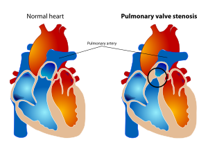
Pulmonary valve stenosis (PVS) is a heart valve disorder. Blood going from the heart to the lungs goes through the pulmonary valve, whose purpose is to prevent blood from flowing back to the heart. In pulmonary valve stenosis this opening is too narrow, leading to a reduction of flow of blood to the lungs.[1][5]
While the most common cause of pulmonary valve stenosis is congenital heart disease, it may also be due to a malignant carcinoid tumor. Both stenosis of the pulmonary artery and pulmonary valve stenosis are forms of pulmonic stenosis (nonvalvular and valvular, respectively)[6] but pulmonary valve stenosis accounts for 80% of pulmonic stenosis. PVS was the key finding that led Jacqueline Noonan to identify the syndrome now called Noonan syndrome.
Symptoms and signs[]

Among some of the symptoms consistent with pulmonary valve stenosis are the following:[2]
- Heart murmur
- Cyanosis
- Dyspnea
- Dizziness
- Upper thorax pain
- Developmental disorders
Cause[]
In regards to the cause of pulmonary valve stenosis a very high percentage are congenital, the right ventricular flow is hindered (or obstructed by this). The cause in turn is divided into: valvular, external and intrinsic (when it is acquired).[3]
Pathophysiology[]
The pathophysiology of pulmonary valve stenosis consists of the valve leaflets becoming too thick (therefore not separate one from another), which can cause high pulmonary pressure, and pulmonary hypertension. This however, does not mean the cause is always congenital.[7]
The left ventricle can be changed physically, these changes are a direct result of right ventricular hypertrophy. Once the obstruction is subdued, it (the left ventricle) can return to normal.[8]
Diagnosis[]

The diagnosis of pulmonary valve stenosis can be achieved via echocardiogram, as well as a variety of other means among them are: ultrasound, in which images of the heart chambers in utero where the tricuspid valve has thickening (or due to Fallot's tetralogy, Noonan's syndrome, and other congenital defects) and in infancy auscultation of the heart can reveal identification of a murmur.[4]
Some other conditions to contemplate (in diagnosis of pulmonic valvular stenosis) are the following:[2]
- Infundibular stenosis
- Supravalvular pulmonary stenosis
- Dysplastic pulmonic valve stenosis
Treatment[]
In terms of treatment for pulmonary valve stenosis, valve replacement or surgical repair (depending upon whether the stenosis is in the valve or vessel) may be indicated. If the valve stenosis is of congenital origin, balloon valvuloplasty is another option, depending on the case. Valves made from animal or human tissue (are used for valve replacement), in adults metal valves can be used.[9][10]
Epidemiology[]
The epidemiology of pulmonary valve stenosis can be summed up by the congenital aspect which is the majority of cases, in broad terms PVS is rare in the general population.[4]
References[]
- ^ a b "Pulmonary valve stenosis: MedlinePlus Medical Encyclopedia". www.nlm.nih.gov. Retrieved 2015-11-18.
- ^ a b c "Pulmonic Valvular Stenosis Clinical Presentation: History, Physical, Causes". emedicine.medscape.com. Retrieved 2015-11-18.
- ^ a b Wang, Andrew; Bashore, Thomas M. (2010-01-14). Valvular Heart Disease. Springer Science & Business Media. p. 266. ISBN 9781597454117.
- ^ a b c "Pulmonary Valve Disease. About Pulmonary valve disease | Patient". Patient. Retrieved 2015-11-18.
- ^ Choices, NHS. "Congenital heart disease - Types - NHS Choices". www.nhs.uk. Retrieved 2015-11-18.
- ^ Ren, XM; et al. (2014-12-23), "Pulmonic stenosis", Medscape.
- ^ Levine, Shel; Coyne, Brian J.; Colvin, Lisa Cooper (2015-02-13). Clinical Exercise Electrocardiography. Jones & Bartlett Publishers. p. 14. ISBN 9781284034202.
- ^ "Valvar Pulmonary Stenosis: Background, Pathophysiology, Epidemiology". 2018-06-07. Cite journal requires
|journal=(help) - ^ Choices, NHS. "Congenital heart disease - Treatment - NHS Choices". www.nhs.uk. Retrieved 2015-11-18.
- ^ "Balloon dilatation of pulmonary valve stenosis | Guidance and guidelines | NICE". www.nice.org.uk. Retrieved 2015-11-18.
Further reading[]
- Ladusans, E J; Qureshi, S A; Parsons, J M; Arab, S; Baker, E J; Tynan, M (1990-06-01). "Balloon dilatation of critical stenosis of the pulmonary valve in neonates". British Heart Journal. 63 (6): 362–367. doi:10.1136/hrt.63.6.362. ISSN 0007-0769. PMC 1024522. PMID 2375899.
- Crocetti, Michael; Barone, Michael A.; Oski, Frank A. (2004-01-01). Oski's Essential Pediatrics. Lippincott Williams & Wilkins. ISBN 9780781737708.
External links[]
| Scholia has a topic profile for Pulmonary valve stenosis. |
- Valvular heart disease