Condyloid fossa
| Condyloid fossa | |
|---|---|
 Occipital bone. Outer surface. (Condyloid fossa visible but not labeled.) | |
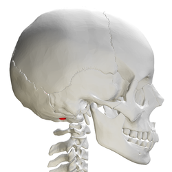 Skull and cervical vertebra. Position of condyloid fossa shown in red. | |
| Details | |
| Identifiers | |
| Latin | Fossa condylaris |
| TA98 | A02.1.04.017 |
| TA2 | 560 |
| FMA | 75310 |
| Anatomical terms of bone | |
Behind either condyle of the lateral parts of occipital bone is a depression, the condyloid fossa (or condylar fossa), which receives the posterior margin of the superior facet of the atlas when the head is bent backward; the floor of this fossa is sometimes perforated by the condyloid canal, through which an emissary vein passes from the transverse sinus.
Additional images[]
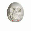
Human skull seen from below. Position of condyloid fossa shown in red.

Skull and cervical vertebra. Position of condyloid fossa shown in red.
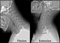
X-ray of cervical spine (neck) in flexion and extension (bending backwards)


See also[]
- Occipital condyle
- Atlas
References[]
![]() This article incorporates text in the public domain from page 131 of the 20th edition of Gray's Anatomy (1918)
This article incorporates text in the public domain from page 131 of the 20th edition of Gray's Anatomy (1918)
External links[]
| Wikimedia Commons has media related to Condyloid fossa. |
| Authority control: Scientific databases |
|---|
This human musculoskeletal system article is a stub. You can help Wikipedia by . |
- Wikipedia articles incorporating text from the 20th edition of Gray's Anatomy (1918)
- Bones of the head and neck
- Musculoskeletal system stubs