Facial canal
| Facial canal | |
|---|---|
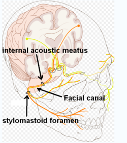 Route of facial nerve, with facial canal labeled | |
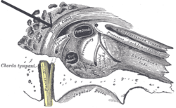 View of the inner wall of the tympanum. (Facial canal visible in upper left; promontory labeled at center) | |
| Details | |
| System | skeletal |
| Nerve | facial nerve (CN VII) |
| Identifiers | |
| Latin | canalis nervi facialis, canalis facialis |
| TA98 | A02.1.06.009 |
| TA2 | 688 |
| FMA | 54952 |
| Anatomical terminology | |
The facial canal (canalis nervi facialis), also known as the Fallopian canal, is a Z-shaped canal running through the temporal bone of the skull. It runs from the internal acoustic meatus to the stylomastoid foramen. It contains the facial nerve (CN VII), after which it is named.
Structure[]
The facial canal runs from the internal auditory meatus to the stylomastoid foramen. In humans it is approximately 3 cm long, which makes it the longest human osseous canal of a nerve.[1] It is located within the middle ear region, according to its shape it is divided into three main segments: the labyrinthine, the tympanic, and the mastoidal segment.[2] It contains the facial nerve (CN VII), after which it is named.[3]
Prominence[]
The prominence of the facial canal (or prominence of the aqueduct of Fallopius) indicates the position of the bony facial canal in which the facial nerve is contained; this canal traverses the medial wall of the tympanic cavity above the oval window, and behind this it curves nearly vertically downward along the posterior wall.
Function[]
The facial canal contains the facial nerve (CN VII), after which it is named.[1]
Clinical significance[]
The facial canal may be interrupted in some people.[1] This may lead to the facial nerve being split into 2 or 3 fibres, or it may be poorly formed or congenitally absent on one side.[1]
History[]
The facial canal was first described by Gabriele Falloppio.[4] This is why it may also be known as the Fallopian canal.[4]
Gallery[]

Lateral head anatomy detail. Facial nerve dissection.

Tympanic cavity. Facial canal. Internal carotid artery.
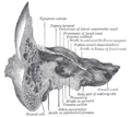
Coronal section of right temporal bone. Prominence of the facial canal labeled at top, fourth from the left.


See also[]
- Facial nerve
- Prominence of the facial canal
- Hiatus of the facial canal
References[]
- ^ a b c d Weiglein AH (June 1996). "Postnatal development of the facial canal. An investigation based on cadaver dissections and computed tomography". Surgical and Radiologic Anatomy. 18 (2): 115–23. doi:10.1007/BF01795229. PMID 8782317. S2CID 25764734.
- ^ Weiglein AH, Anderhuber W, Jakse R, Einspieler R (1994). "Imaging of the facial canal by means of multiplanar angulated 2-D-high-resolution CT-reconstruction". Surgical and Radiologic Anatomy. 16 (4): 423–427. doi:10.1007/BF01627665. PMID 7725200. S2CID 10477288.
- ^ Nager, George T.; Proctor, Bruce (1991-06-01). "Anatomie Variations and Anomalies Involving the Facial Canal". Otolaryngologic Clinics of North America. 24 (3): 531–553. doi:10.1016/S0030-6665(20)31114-2. ISSN 0030-6665.
- ^ a b Abing W, Rauchfuss A (2005). "Fetal development of the tympanic part of the facial canal". European Archives of Oto-Rhino-Laryngology. 243 (6): 374–377. doi:10.1007/bf00464645. PMID 3566620. S2CID 12712839.
| Authority control: Scientific databases |
|---|
- Foramina of the skull
- Ear