Middle nasal concha
| Middle nasal concha | |
|---|---|
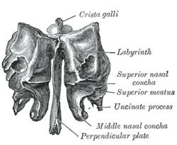 Ethmoid bone from behind. | |
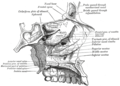
 Lateral wall of nasal cavity, showing ethmoid bone in position. (Middle nasal concha is at bottom of pink region.) | |
| Details | |
| Identifiers | |
| Latin | Concha media |
| TA98 | A06.1.02.014 |
| TA2 | 735 |
| FMA | 57459 |
| Anatomical terms of bone | |
The medial surface of the labyrinth of ethmoid consists of a thin lamella, which descends from the under surface of the cribriform plate, and ends below in a free, convoluted margin, the middle nasal concha (middle nasal turbinate).
It is rough, and marked above by numerous grooves, directed nearly vertically downward from the cribriform plate; they lodge branches of the olfactory nerves, which are distributed to the mucous membrane covering the superior nasal concha.
Additional images[]

Nose and nasal cavities

Ethmoid bone from the right side.
Roof, floor, and lateral wall of left nasal cavity.
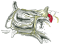
The sphenopalatine ganglion and its branches.

Coronal section of nasal cavities.

Sagittal section of nose, mouth, pharynx, and larynx.

Nasal conchae


See also[]
- Nasal concha
References[]
![]() This article incorporates text in the public domain from page 156 of the 20th edition of Gray's Anatomy (1918)
This article incorporates text in the public domain from page 156 of the 20th edition of Gray's Anatomy (1918)
External links[]
- Anatomy figure: 33:01-01 at Human Anatomy Online, SUNY Downstate Medical Center
- Anatomy figure: 22:02-10 at Human Anatomy Online, SUNY Downstate Medical Center
- upstate.edu - Frontal
- Atlas image: rsa1p9 at the University of Michigan Health System - lateral
- Atlas image: rsa1p6 at the University of Michigan Health System - coronal
- "Anatomy diagram: 34256.000-1". Roche Lexicon - illustrated navigator. Elsevier. Archived from the original on 2012-12-27.
| Authority control: Scientific databases |
|---|
This human musculoskeletal system article is a stub. You can help Wikipedia by . |
- Wikipedia articles incorporating text from the 20th edition of Gray's Anatomy (1918)
- Bones of the head and neck
- Musculoskeletal system stubs